From Awareness and Diagnosis to Lumpectomy, Mastectomy and Beyond
Richard Purchon FRGS AC RODP
Introduction
During my Operating Department Practitioner training, I spent a considerable amount of time working as a scrub practitioner with Mr Haresh Devalia, Consultant Breast and Oncoplastic Surgeon at Maidstone and Tunbridge Wells NHS Trust.
Mr Devalia developed extensive experience in breast oncology, breast-conserving and oncoplastic surgery, mastectomy and reconstructive techniques. Working alongside him exposed me to a broad spectrum of breast cancer surgery and gave me an early appreciation that breast cancer surgery is considerably more complex than the commonly used distinction between a “lumpectomy” and a “mastectomy”.
At the end of that period I was offered the opportunity to join his team. It was an attractive prospect, but I ultimately chose a different direction and moved into cardiac anaesthesia, which went on to form a major part of my clinical career.
The experience nevertheless stayed with me.
Breast cancer surgery is not one operation. It represents a spectrum ranging from the removal of a relatively small volume of breast tissue through lymph-node staging and mastectomy to microsurgical reconstruction and, occasionally, major thoracic and chest-wall surgery.
Just as importantly, the operation itself represents only one point in a much longer patient journey.
For some patients that journey begins with an abnormality identified during screening. For others it begins while showering, dressing or simply noticing that something about their breast or chest feels different.
The pathway may subsequently involve:
awareness → referral → imaging → biopsy → multidisciplinary review → localisation → surgery → lymph-node assessment → histology → further treatment → recovery and follow-up.
This article follows that journey from the perspective of the qualified healthcare practitioner, with particular emphasis on the information relevant to nurses, Operating Department Practitioners and other members of the perioperative team.
The Breast Cancer Patient Journey
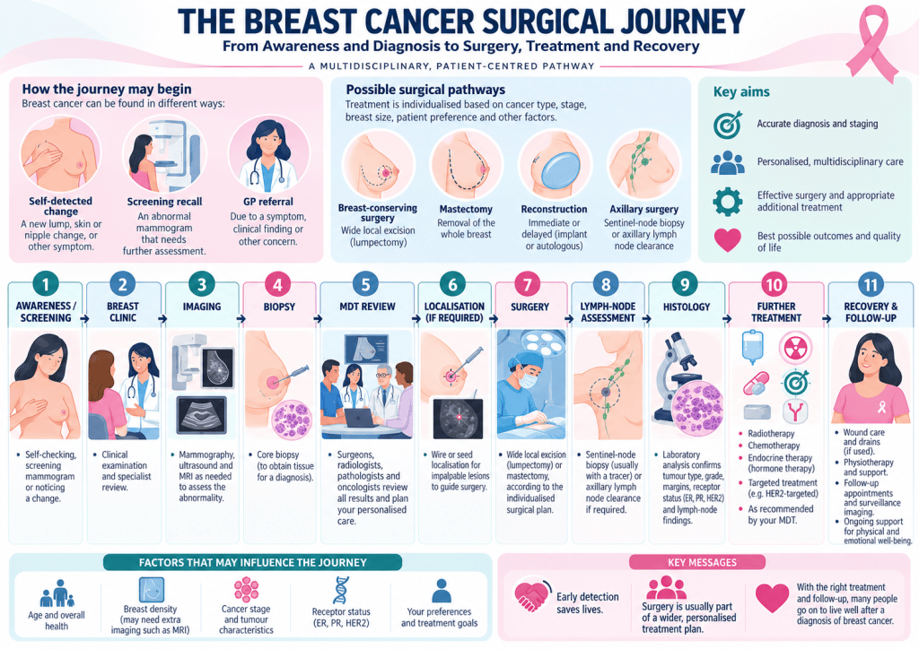
(IMAGE 1 — THE BREAST CANCER SURGICAL JOURNEY)
The journey diagram provides the framework for the whole article.
It is important to recognise that no two patients necessarily follow exactly the same pathway.
A screen-detected lesion may be clinically impalpable and require image-guided localisation before surgery.
A patient presenting with a palpable mass may require no localisation at all.
Some patients undergo breast-conserving surgery.
Others require mastectomy.
Some undergo sentinel lymph-node biopsy, whereas others already have proven nodal disease and require a different axillary strategy.
Reconstruction may be immediate, delayed or not undertaken.
The treatment pathway is therefore individualised according to:
- tumour pathology
- tumour size
- disease distribution
- lymph-node status
- receptor status
- previous treatment
- genetic risk
- comorbidities
- anatomy
- patient preference.
The pathway is multidisciplinary from the beginning.
Stage 1: Awareness and Recognition
Knowing What Is Normal
For many patients, their cancer journey begins because they recognise something that is not normal for them.
This is where organisations such as CoppaFeel! have made an important contribution to breast and chest awareness, particularly among younger people.
The objective of breast awareness is not simply to repeatedly search for a lump.
It is to become familiar with one’s own breast or chest and recognise change.
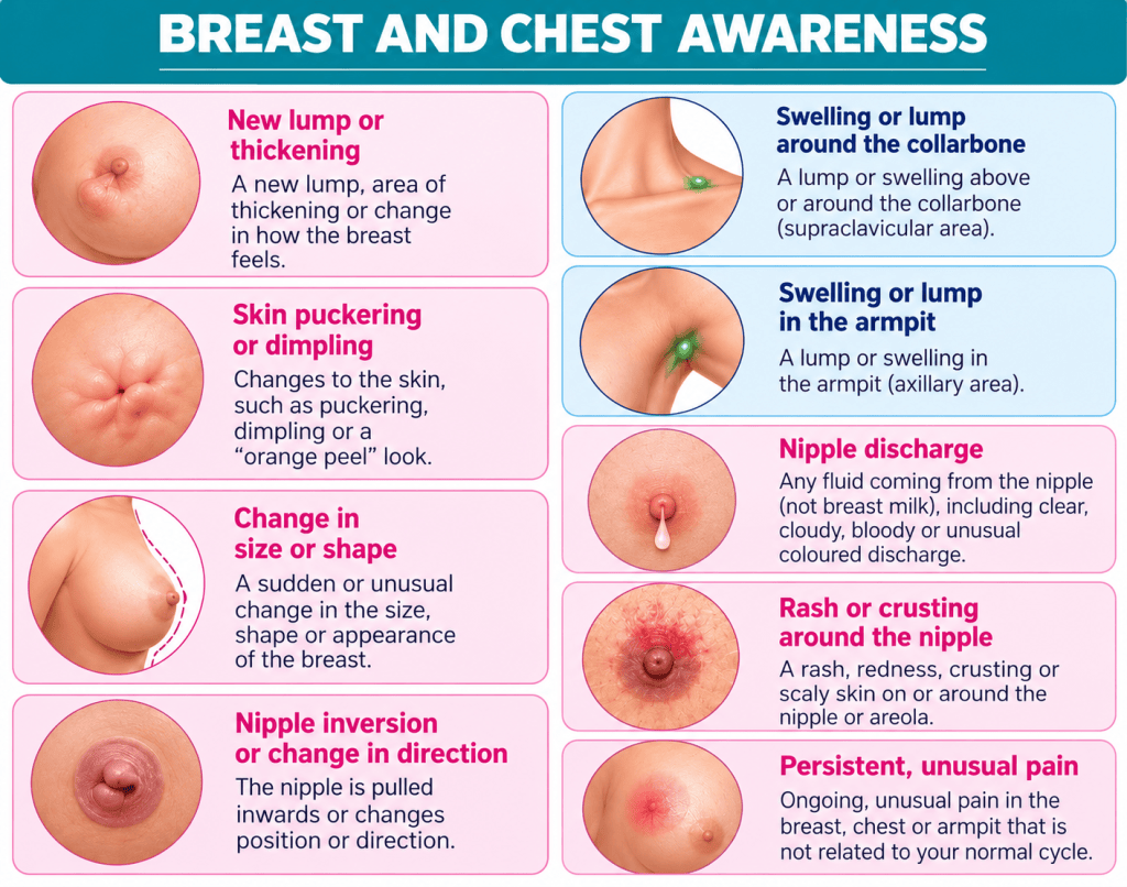
(IMAGE 2 — BREAST AND CHEST AWARENESS)
Changes that may warrant clinical assessment include:
- a new lump or thickening
- swelling in the breast or axilla
- swelling around the clavicle
- skin puckering or dimpling
- peau d’orange-type skin changes
- nipple inversion or change in direction
- unexpected nipple discharge
- rash or crusting around the nipple
- an unexplained change in breast size or contour
- persistent unusual breast, chest-wall or axillary discomfort.
None of these findings automatically means cancer.
Their importance lies in recognising a new or unexplained change and seeking appropriate clinical assessment.
For the theatre practitioner this provides useful context.
By the time somebody appears on an operating list, they may already have experienced several weeks of uncertainty, examinations, imaging, biopsy and difficult conversations.
The operation may be familiar to the staff.
For the patient it is rarely routine.
Stage 2: The Breast Clinic
Once an abnormality has been identified, assessment is directed towards establishing what it represents.
This commonly involves the principles of triple assessment:
Clinical assessment
History and examination of the:
- breast
- nipple
- overlying skin
- axilla
- supraclavicular region where appropriate.
Imaging
Depending upon age, presentation and previous investigations, this may involve:
- mammography
- ultrasound
- additional mammographic views
- MRI in selected patients.
Tissue diagnosis
Suspicious abnormalities usually require tissue sampling.
Core biopsy allows tissue architecture to be examined and may provide information about tumour type and biological characteristics.
The important point is that clinical examination, imaging and pathology are interpreted together, not in isolation.
Stage 3: Understanding the Anatomy
Before considering breast surgery it is useful to understand the anatomical layers through which the surgeon operates.
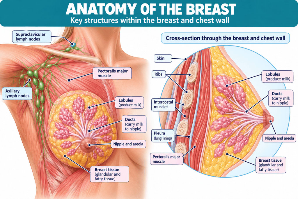
(IMAGE 3 — ANATOMY OF THE BREAST)
The breast consists of:
- glandular lobules
- ducts
- adipose tissue
- connective tissue
- skin
- nipple-areolar complex.
Posteriorly, the breast lies predominantly over pectoralis major, with the ribs, intercostal muscles and pleural cavity deeper still.
The relationship can therefore be simplified as:
skin
↓
breast tissue
↓
pectoral fascia / pectoralis major
↓
ribs and intercostal muscles
↓
parietal pleura
↓
lung
This relationship becomes increasingly important as breast cancer surgery progresses from straightforward breast-conserving surgery towards mastectomy, reconstruction and, occasionally, full-thickness chest-wall resection.
Stage 4: Breast Lymphatic Drainage
The breast cannot be considered independently from its lymphatic drainage.
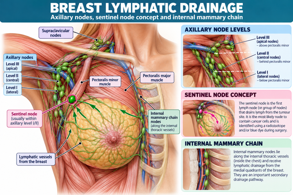
(IMAGE 4 — BREAST LYMPHATIC DRAINAGE)
Much of the breast drains towards the axillary lymph nodes.
Additional drainage can occur towards:
- internal mammary nodes
- supraclavicular nodes
- other regional lymphatic pathways.
The axillary nodes are conventionally described in relation to pectoralis minor.
Broadly:
- Level I nodes lie lateral/inferior to pectoralis minor.
- Level II nodes lie posterior to pectoralis minor.
- Level III nodes lie medial/superior to it towards the axillary apex.
This anatomical organisation matters because extent of nodal involvement influences staging and management.
Stage 5: The Sentinel Lymph Node
The sentinel lymph node is the first lymph node—or group of nodes—receiving lymphatic drainage from a particular tumour-bearing region.
Conceptually:
breast tumour
↓
lymphatic channel
↓
sentinel node
↓
additional regional nodes
If tumour cells begin spreading through the lymphatic system, the sentinel node is among the most likely places for them to be identified.
This permits selected patients to undergo sentinel-node biopsy rather than automatically undergoing extensive axillary clearance.
Stage 6: Surgical Planning
Once imaging and pathology have been reviewed, treatment is discussed within the multidisciplinary team.
The operation may involve:
- wide local excision
- oncoplastic breast-conserving surgery
- mastectomy
- sentinel lymph-node biopsy
- targeted axillary surgery
- axillary lymph-node clearance
- reconstruction
- combinations of the above.
Tumour biology may also determine whether systemic treatment occurs before surgery.
The term breast cancer surgery therefore covers a remarkably wide range of procedures.
Stage 7: Wide Local Excision — Lumpectomy
The commonly understood term lumpectomy is frequently described surgically as a wide local excision — WLE.
The purpose is not simply to remove the visible lump.
The surgeon removes the tumour together with surrounding breast tissue to allow pathological assessment of the resection margins.
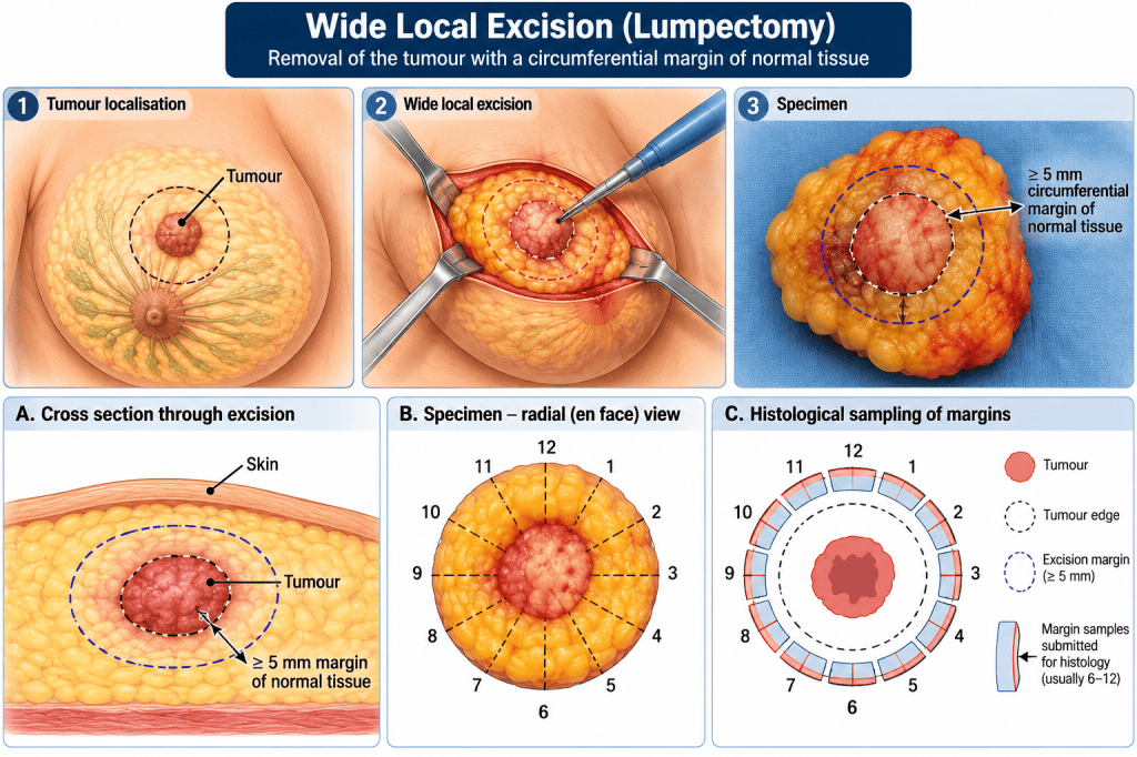
(IMAGE 5 — WIDE LOCAL EXCISION / LUMPECTOMY)
The principle is:
tumour + surrounding normal breast tissue → oriented surgical specimen → histopathological margin assessment.
A useful distinction must be made between the gross surgical margin illustrated in diagrams and the final pathological definition of an adequate margin.
The surgeon does not simply measure an arbitrary number of millimetres around every tumour and declare the operation complete.
Margin adequacy depends upon the underlying pathology, microscopic assessment and current oncological guidance.
The illustration should therefore be understood as demonstrating the principle of circumferential removal of apparently normal tissue around the tumour, rather than prescribing one universal margin measurement.
Stage 8: Why Margins Matter
The surgeon can see and palpate tissues.
The surgeon cannot reliably see microscopic tumour cells.
Consequently, final pathology may demonstrate disease closer to a margin than expected.
This may result in:
- no further breast surgery
- cavity-margin re-excision
- further wide local excision
- conversion to mastectomy in selected cases.
A patient returning for another operation following a WLE has therefore not necessarily experienced a technical failure.
New information has become available following microscopic assessment of the specimen.
This is an important distinction when communicating with patients.
Stage 9: The Impalpable Lesion
Not all breast cancers can be felt.
Screening frequently detects:
- small lesions
- distortions
- microcalcification
- biopsy-proven abnormalities that remain clinically impalpable.
The surgeon therefore requires a means of locating something that cannot reliably be identified by touch.
This is where image-guided localisation becomes important.
Stage 10: Wire-Guided Localisation
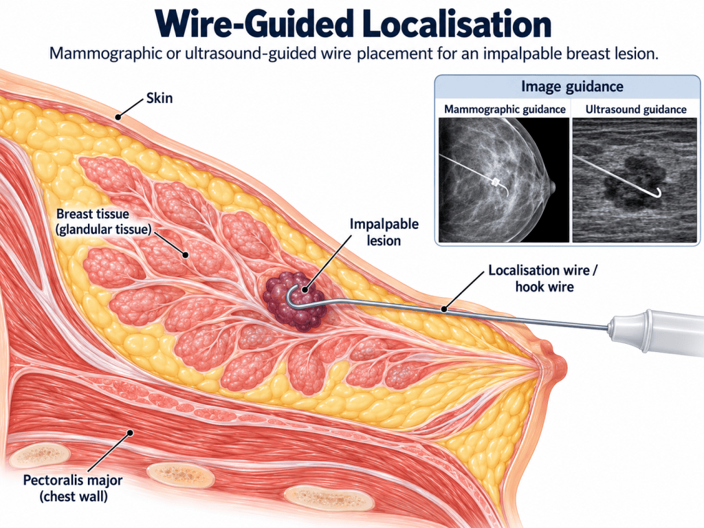
(IMAGE 6 — WIRE-GUIDED LOCALISATION)
Using mammographic or ultrasound guidance, a localisation wire can be introduced so that its tip lies within or immediately adjacent to the target lesion.
The patient subsequently arrives in theatre with part of the wire remaining external to the breast and secured with a dressing.
From the perioperative perspective, this creates several practical considerations.
The localisation wire must be protected during:
- patient transfer
- removal or adjustment of clothing
- positioning
- skin preparation
- application of surgical drapes.
Accidental displacement could compromise localisation.
The presence, site and integrity of the localisation device therefore become part of the preoperative checking process.
Stage 11: Wire-Guided Surgical Excision
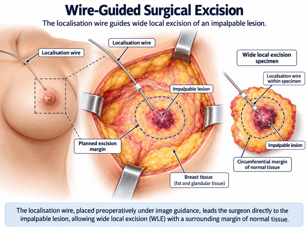
(IMAGE 7 — WIRE-GUIDED SURGICAL EXCISION)
The wire provides a three-dimensional guide towards the lesion.
It does not merely indicate where to make the skin incision.
The surgeon follows the trajectory of the wire towards the target and removes the relevant volume of tissue with an oncologically appropriate margin.
The wire is generally removed with the specimen.
The specimen must then be:
- correctly orientated
- labelled
- documented
- transferred safely for any required imaging and subsequent pathology.
For an impalpable lesion, this is particularly important because the surgeon cannot simply look at the specimen and be certain that the original radiological target has been removed.
Stage 12: Specimen Imaging
Some specimens—particularly those containing mammographically detected microcalcification, biopsy clips or localisation devices—may undergo specimen radiography.
This asks an extremely practical question:
Is the radiological target actually inside the piece of tissue that has been removed?
The image may demonstrate:
- microcalcification
- biopsy clip
- guide wire
- seed marker
- relationship of the abnormality to the specimen margins.
If the target is not appropriately represented, additional tissue may sometimes be removed before the patient leaves theatre.
This demonstrates the close relationship between:
radiology → surgery → pathology.
Stage 13: Seed Localisation
Wire localisation is not the only option.
Small markers can also be placed into an impalpable lesion before surgery.
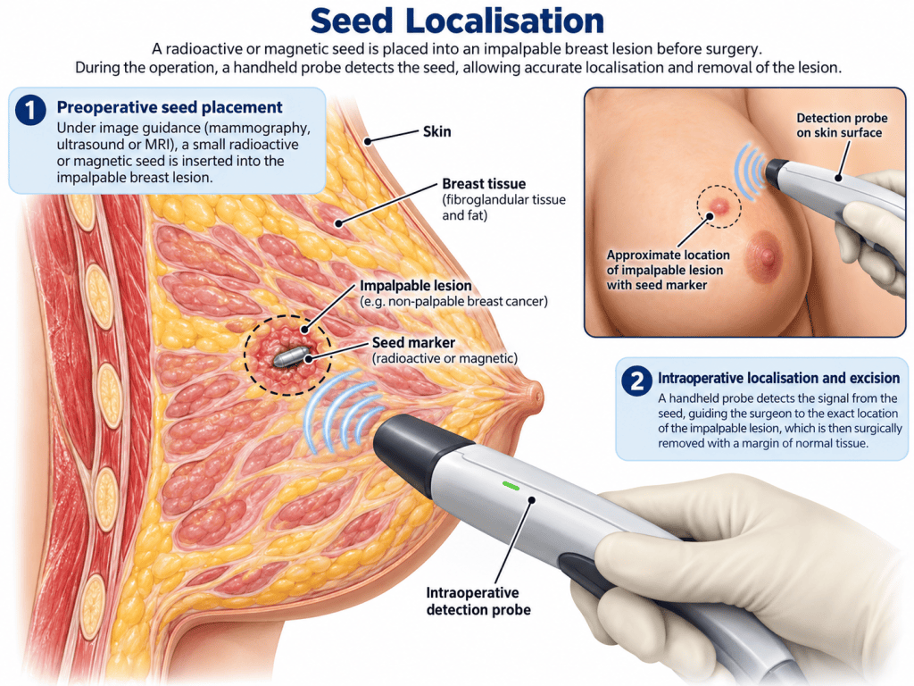
(IMAGE 8 — SEED LOCALISATION)
Depending upon the technology used, the marker may be:
- radioactive
- magnetic
- reflector-based
- detectable using another dedicated system.
The broad principle is the same:
marker placed under image guidance
↓
marker detected intraoperatively
↓
surgeon localises lesion
↓
lesion and marker removed within specimen.
Unlike a protruding wire, some seed-based systems allow localisation to occur before the day of surgery, potentially simplifying theatre-day logistics.
However, the practitioner needs to know which localisation system is being used, because the detection technology and equipment are not interchangeable.
Stage 14: Sentinel Lymph-Node Mapping with Radioisotope
Localisation of a breast lesion and localisation of a sentinel lymph node are two different procedures.
Sentinel-node mapping follows lymphatic drainage rather than identifying the primary tumour itself.
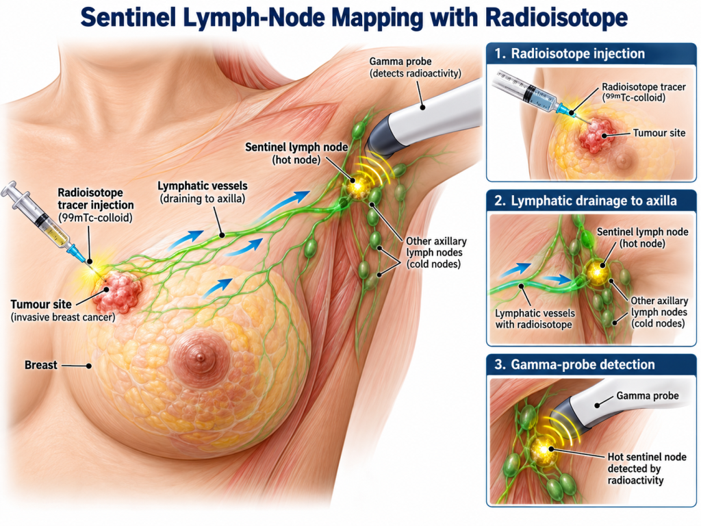
(IMAGE 9 — SENTINEL LYMPH-NODE MAPPING WITH RADIOISOTOPE)
A small quantity of radioactive tracer is injected according to the local protocol.
The tracer enters the lymphatic channels and travels towards the draining nodes.
The sentinel node becomes relatively radioactive or “hot”.
In theatre a handheld gamma probe allows the surgeon to detect radioactive activity.
As the probe approaches the sentinel node, the count rises.
The gamma probe therefore converts an invisible physiological pathway into information that can be heard and measured.
For the theatre practitioner, important considerations include:
- availability of the correct probe
- correct equipment setup
- checking probe function
- awareness of local radioactive-material procedures
- communication regarding specimen handling.
Stage 15: Blue-Dye Sentinel-Node Mapping
Another method of identifying lymphatic drainage uses visible dye.
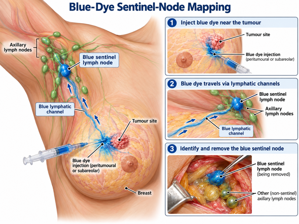
(IMAGE 10 — BLUE-DYE SENTINEL-NODE MAPPING)
The blue dye enters lymphatic channels and may allow the surgeon to see:
blue lymphatic vessel → blue sentinel node.
The dye and radioisotope can therefore provide two different methods of identifying the same anatomical destination.
The isotope produces a measurable radioactive signal.
The dye produces a visible pathway.
Use varies according to local practice and technique.
Anaesthetic implications
Blue dye is not simply a surgical issue.
Significant hypersensitivity reactions, including anaphylaxis, can occur.
Administration should therefore be communicated clearly to the anaesthetic team.
Depending on the agent used, temporary skin staining, urine discolouration and effects on optical monitoring may also be observed.
This demonstrates an important perioperative principle:
Something injected by the surgeon may immediately become relevant to the anaesthetic practitioner.
Stage 16: Axillary Surgical Anatomy
Where more extensive axillary surgery is required, the anatomy becomes substantially more complex.
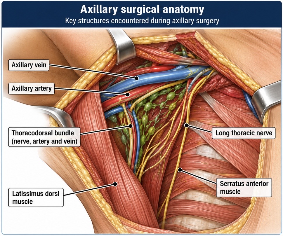
(IMAGE 11 — AXILLARY SURGICAL ANATOMY)
Important structures include:
- axillary vein
- axillary artery
- long thoracic nerve
- thoracodorsal nerve
- thoracodorsal artery and vein
- latissimus dorsi
- serratus anterior.
The long thoracic nerve runs in relation to serratus anterior.
Damage may result in impaired serratus function and scapular winging.
The thoracodorsal bundle supplies latissimus dorsi and is also important when that muscle is considered for reconstruction.
The intercostobrachial nerves are another relevant structure because disturbance can contribute to postoperative numbness or neuropathic symptoms.
Stage 17: Axillary Lymph-Node Clearance
An axillary clearance represents a considerably more extensive dissection than sentinel-node biopsy.
Potential postoperative consequences include:
- seroma
- altered sensation
- neuropathic pain
- shoulder stiffness
- lymphoedema
- wound complications
- nerve injury.
This is why modern breast cancer surgery aims to obtain the information needed for staging while avoiding unnecessary disruption of normal lymphatic anatomy where possible.
Stage 18: When Breast Conservation Is Not Appropriate
Breast-conserving surgery may not always provide an appropriate oncological or cosmetic outcome.
Reasons for recommending mastectomy may include:
- tumour size relative to breast size
- multicentric disease
- extensive DCIS
- inability to obtain satisfactory margins
- recurrence
- previous treatment
- genetic considerations
- patient preference.
Mastectomy should not simply be thought of as a larger lumpectomy.
It represents a different surgical strategy.
Stage 19: Mastectomy Anatomy
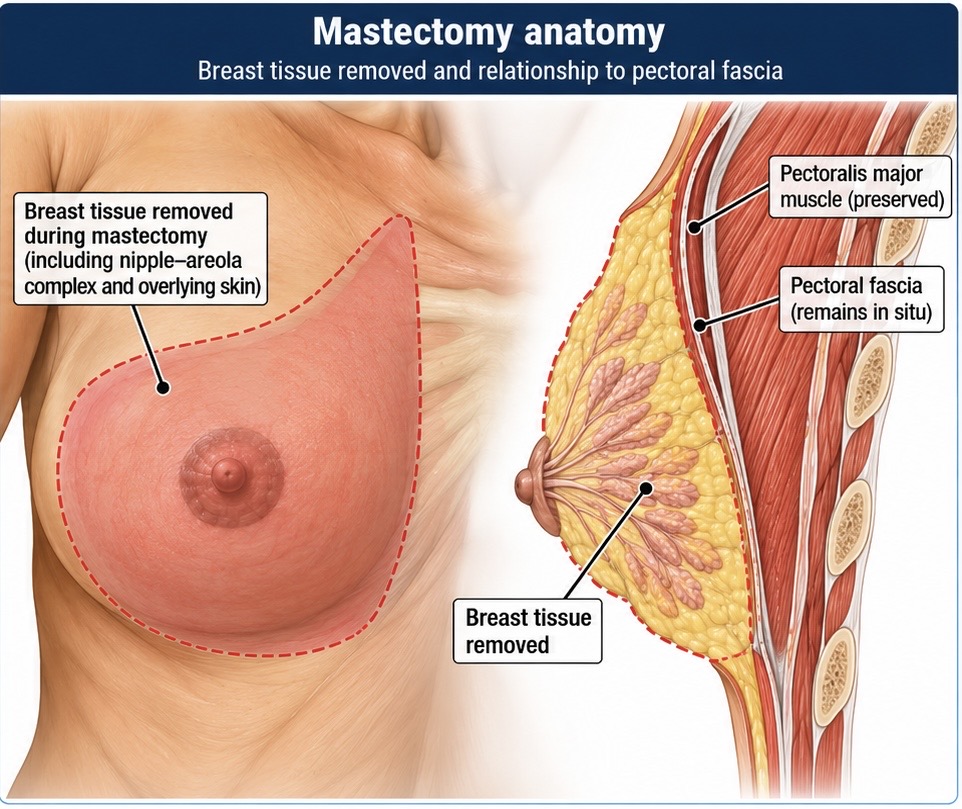
(IMAGE 12 — MASTECTOMY ANATOMY)
During mastectomy, breast tissue is removed from beneath the skin envelope and from the plane overlying pectoralis major.
Depending upon the operation this may include:
- nipple-areolar complex
- varying amounts of skin
- almost all macroscopic breast tissue.
The relationship with the pectoral fascia is clinically important.
Different operative techniques and oncological circumstances may influence how fascia is managed; therefore diagrams showing fascia preservation should be understood as illustrating the relevant tissue planes rather than defining every possible mastectomy technique.
The underlying pectoralis major muscle is usually preserved unless directly involved by disease or deliberately used within another reconstructive strategy.
Stage 20: Skin-Sparing and Nipple-Sparing Surgery
Modern mastectomy does not always require removal of the entire visible breast skin envelope.
Skin-sparing mastectomy
Much of the skin is retained to facilitate reconstruction.
Nipple-sparing mastectomy
Where oncologically appropriate, the nipple-areolar complex can also be preserved.
The essential point is:
preserving skin does not mean preserving the breast tissue beneath it.
The oncological component and reconstructive component remain separate considerations.
Stage 21: Breast Reconstruction
Breast reconstruction may be:
- immediate
- delayed
- implant based
- autologous
- combined.
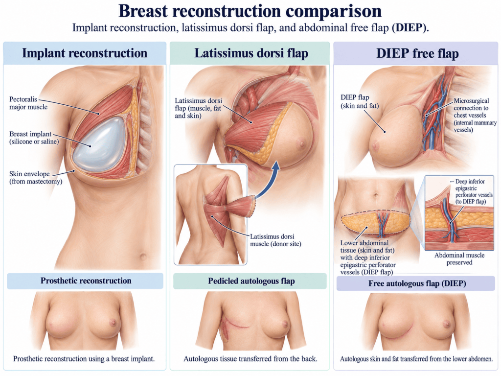
(IMAGE 13 — BREAST RECONSTRUCTION COMPARISON)
Three broad examples illustrate the increasing complexity.
Implant Reconstruction
A prosthetic implant is used to recreate breast volume.
This may involve:
- direct-to-implant reconstruction
- staged tissue-expander reconstruction
- prepectoral or subpectoral positioning depending upon technique.
The perioperative practitioner needs to consider:
- implant documentation
- antibiotic policy
- sterile handling
- specialist equipment
- drains where used.
Latissimus Dorsi Flap
The latissimus dorsi muscle, with associated skin and fat where required, can be mobilised from the back and transferred to the breast.
Its vascular supply through the thoracodorsal system makes knowledge of axillary anatomy especially relevant.
The flap may be used alone or in combination with an implant.
DIEP Free Flap
The deep inferior epigastric perforator flap uses abdominal skin and fat while attempting to preserve the rectus muscle.
Unlike a pedicled flap, the tissue is completely disconnected from its original circulation and reconnected microsurgically to recipient vessels in the chest.
This transforms the procedure into prolonged microsurgery with implications for:
- operating duration
- warming
- pressure-area protection
- vascular access
- thromboprophylaxis
- fluid management
- flap monitoring
- postoperative destination.
Stage 22: When Breast Cancer Reaches the Chest Wall
Most breast cancer surgery remains outside the thoracic cavity.
Locally advanced disease or recurrence may occasionally extend deeper.
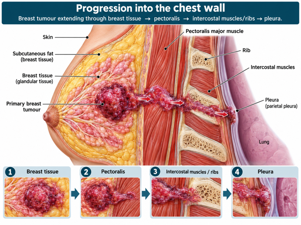
(IMAGE 14 — PROGRESSION INTO THE CHEST WALL)
The potential anatomical progression is:
breast tissue
↓
pectoral fascia
↓
pectoralis major
↓
intercostal muscles
↓
ribs / sternum
↓
parietal pleura
↓
intrathoracic structures.
This is the point at which the artificial boundary between “breast surgery” and “thoracic surgery” becomes increasingly blurred.
The patient may require involvement from:
- breast surgeons
- thoracic surgeons
- plastic/reconstructive surgeons
- anaesthetists familiar with major thoracic surgery
- critical-care teams.
Stage 23: Full-Thickness Chest-Wall Resection
When tumour involves ribs or sternum, obtaining adequate oncological clearance may require full-thickness chest-wall resection.
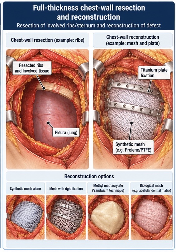
(IMAGE 15 — FULL-THICKNESS CHEST-WALL RESECTION AND RECONSTRUCTION)
This can involve removal of:
- skin
- subcutaneous tissue
- muscle
- one or more ribs
- parts of the sternum
- involved pleura.
The resulting defect may compromise:
- chest-wall stability
- respiratory mechanics
- protection of underlying structures.
Consequently, reconstruction may be required.
Options include combinations of:
- synthetic mesh
- biological mesh
- rigid fixation
- plates
- methyl-methacrylate-based techniques
- muscle or myocutaneous flap coverage.
If the pleural cavity is entered, chest drainage may be required.
If pulmonary resection is necessary, the anaesthetic plan may progress into true thoracic anaesthesia, potentially including lung-isolation techniques.
At this point, the operation bears little resemblance to a conventional lumpectomy.
Yet it represents the far end of the same breast cancer surgical spectrum.
Stage 24: The Theatre Pathway
The whole perioperative journey can be considered as a linked sequence rather than a collection of isolated tasks.
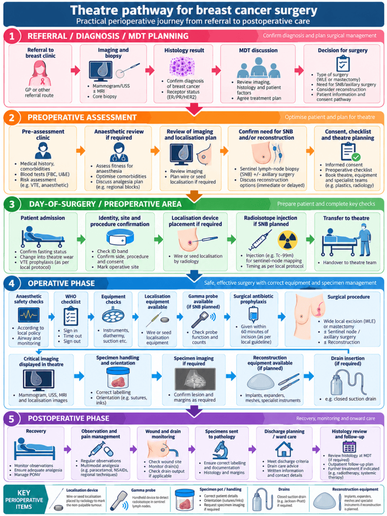
(IMAGE 16 — THEATRE PATHWAY FOR BREAST CANCER SURGERY)
A procedure listed simply as:
WLE + SLNB
may actually involve:
preoperative imaging
↓
localisation device
↓
radioisotope injection
↓
anaesthetic assessment
↓
WHO checklist
↓
gamma-probe preparation
↓
wide local excision
↓
sentinel-node biopsy
↓
specimen orientation
↓
specimen imaging
↓
pathology
↓
postoperative recovery.
Similarly, a listing of:
mastectomy + reconstruction
may represent many hours of multidisciplinary surgery involving several specialist teams.
Stage 25: The ODP and Theatre-Practitioner Perspective
Understanding the patient journey allows the perioperative practitioner to anticipate rather than simply react.
Important questions include:
Has the lesion been localised?
If yes:
- wire?
- radioactive seed?
- magnetic seed?
- another marker?
Is sentinel-node mapping planned?
If yes:
- radioisotope?
- blue dye?
- gamma probe required?
- radioactive specimen handling considerations?
What breast procedure is planned?
- WLE
- oncoplastic excision
- mastectomy
- skin-sparing mastectomy
- nipple-sparing mastectomy.
Is axillary surgery planned?
- sentinel-node biopsy
- targeted axillary surgery
- clearance.
Is reconstruction planned?
- implant
- expander
- latissimus flap
- free flap
- chest-wall reconstruction.
What specimens will be produced?
The practitioner may need to anticipate:
- breast specimen
- separately labelled cavity margins
- sentinel nodes
- axillary contents
- reconstruction tissue
- localisation devices.
Correct orientation and labelling become fundamental.
Stage 26: Specimen Handling
Breast cancer surgery places particular importance on specimen orientation.
The pathologist may need to identify:
- superior
- inferior
- medial
- lateral
- anterior
- posterior margins.
Orientation may use:
- sutures
- clips
- inks
- pins
- diagrams
- local specimen protocols.
The circulating and scrub practitioners should understand the local system rather than assuming that every surgeon uses the same method.
If specimen radiography is required, the chain of communication between theatre and radiology must also be clear.
Stage 27: Drains
Drains are not required following every breast operation.
Where used, they may follow:
- mastectomy
- axillary clearance
- reconstruction
- large tissue dissection
- flap procedures.
Postoperative management includes:
- securing the drain
- recording output
- recognising sudden increases or changes
- monitoring for seroma
- ensuring appropriate patient education before discharge where drains remain in situ.
Stage 28: Recovery
Recovery varies enormously according to the procedure.
A patient following straightforward breast-conserving surgery may return home comparatively quickly.
A patient following free-flap reconstruction or chest-wall resection may require significantly greater postoperative monitoring.
Potential postoperative concerns include:
- pain
- nausea
- wound complications
- haematoma
- seroma
- infection
- sensory disturbance
- reduced shoulder movement
- lymphoedema
- reconstructive failure
- flap perfusion problems.
The operation itself may be complete, but the wider treatment pathway often continues.
Stage 29: Histology and the Next Decision
Definitive histology may provide information regarding:
- tumour size
- histological type
- tumour grade
- margins
- lymph-node status
- hormone receptors
- HER2 status
- other biological markers.
This information returns to the multidisciplinary team.
The next stage may involve:
- no further surgery
- re-excision
- additional axillary treatment
- radiotherapy
- endocrine therapy
- chemotherapy
- targeted therapy.
The surgical journey therefore feeds directly into the wider oncological pathway.
The Patient Behind the Procedure
Clinical pathways divide treatment into departments:
radiology
pathology
surgery
anaesthesia
oncology
physiotherapy
follow-up.
The patient experiences none of these as separate journeys.
They experience one.
A patient undergoing wire-guided WLE may:
- already know they have cancer
- attend radiology while awake
- have a wire placed into the breast
- transfer to theatre
- undergo general anaesthesia
- have the lesion removed
- wait for specimen imaging
- recover from surgery
- then wait several more daysi for definitive histology.
Another patient may wake after mastectomy facing not only postoperative pain but an altered body image, drains and uncertainty about lymph-node results.
A third may undergo many hours of combined oncological and reconstructive surgery before beginning prolonged recovery.
Understanding where the patient is within that journey can change the way apparently small interactions are approached.
Returning to the Beginning
For all the sophistication of modern breast cancer surgery—image-guided wires, radioactive tracers, gamma probes, microsurgery, implants and chest-wall reconstruction—the pathway still begins with recognition.
Something is found.
Sometimes by screening.
Sometimes by a clinician.
Sometimes by the patient.
This is why breast and chest awareness remains important.
Organisations such as CoppaFeel! encourage people to become familiar with what is normal for them and to seek assessment when something changes.
For healthcare practitioners working at the surgical end of that pathway, it provides an important reminder.
Before there was an operating list, there was a person who noticed something was different.
Know what is normal for you.
Look. Feel. Check.
If something changes, get it checked.
The Journey in Summary
Awareness / Screening
↓
Referral to Breast Clinic
↓
Clinical Assessment
↓
Imaging
↓
Biopsy
↓
Pathology
↓
MDT Discussion
↓
Surgical Planning
↓
Wire / Seed Localisation if Required
↓
Wide Local Excision or Mastectomy
↓
Sentinel-Node / Axillary Surgery
↓
Reconstruction Where Appropriate
↓
Specimen Analysis
↓
Definitive Histology
↓
Further Oncology Treatment
↓
Recovery and Follow-Up
Images Used in This Article
- The Breast Cancer Surgical Journey
- Breast and Chest Awareness
- Anatomy of the Breast
- Breast Lymphatic Drainage
- Wide Local Excision / Lumpectomy
- Wire-Guided Localisation
- Wire-Guided Surgical Excision
- Seed Localisation
- Sentinel Lymph-Node Mapping with Radioisotope
- Blue-Dye Sentinel-Node Mapping
- Axillary Surgical Anatomy
- Mastectomy Anatomy
- Breast Reconstruction Comparison
- Progression into the Chest Wall
- Full-Thickness Chest-Wall Resection and Reconstruction
- Theatre Pathway for Breast Cancer Surgery
Suggested Hashtags
#CoppaFeel #CheckYourChest #BreastCancerAwareness #BreastCancer #EarlyDetection #BreastSurgery #BreastCancerSurgery #PatientJourney #Lumpectomy #WideLocalExcision #SentinelLymphNode #Mastectomy #OncoplasticSurgery #BreastReconstruction #ThoracicSurgery #PerioperativeCare #OperatingDepartmentPractitioner #ODP #TheatreNursing #CancerCare #HealthcareEducation

