The Other Side of the Drapes: When the Orthopaedic ODP Becomes the Patient
I had spent years understanding risk.
Professionally, I was an experienced Orthopaedic and cardiothoracic Operating Department Practitioner. I had scrubbed for more tibial intramedullary nails than I could possibly remember and had prepared the anaesthetic requirements for equally many orthopaedic trauma cases.
I knew the implants.
I knew the guidewires.
I knew the reamers.
I knew the locking screws.
I knew the anaesthetic room, the airway equipment, the monitoring, the positioning and the choreography required to get an injured patient safely through a tibial nail.
Outside theatre, I was hardly inexperienced either.
I had spent many years climbing and mountaineering throughout England, Scotland, Wales and the European Alps. I became a Fellow of the Royal Geographical Society with IBG, a member of the Alpine Club and the British Mountain Medicine Society, a Scout climbing instructor and Mountain Leader.
Navigation, weather, terrain, equipment, movement and risk assessment were familiar territory.
And yet none of that prevented one ridiculous accident.
It did not happen on an Alpine face.
It did not happen on an exposed ridge.
It did not happen during a technical climb.
It happened on the walk out.
One Step
It was 18 March 2018.
I had spent the day teaching navigation in North Wales. The day was effectively over and we had approximately two miles left to walk back to the road.
I stepped onto some flattened wet grass.
Hidden beneath it was a large stone.
My foot slipped.
The boot caught.
The combined weight of myself, my gear and my forward momentum continued to carry me onwards while my lower leg twisted backwards beneath me.
Then came two enormous cracks.
Not little pops.
Two explosive reports, almost like gunshots.
I became airborne as my body rotated over the trapped leg before landing heavily on my left side and back. Fortunately, my rucksack absorbed much of the impact.
My right leg, however, was comprehensively broken.
Any attempt to move produced excruciating pain.
I knew immediately what had happened.
There was no period of wondering whether it was “just a sprain”.
I had spent too many years looking at fractures.
I knew.
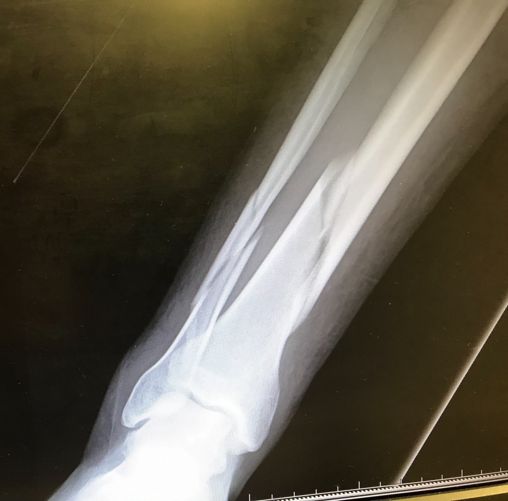
Later radiographs confirmed a displaced spiral/long-oblique fracture of my right tibia with an associated fibular fracture, exactly the kind of pattern one would expect from a substantial torsional mechanism.
And that injury has left me with two questions I still cannot answer with certainty.
I had been wearing Salomon Super Mountain 9 boots: extremely stiff and supportive B2-rated mountain boots.
Would I have sustained such a catastrophic torsional injury had I been wearing a softer, more flexible B0 boot?
Or did the rigidity of the B2 boot, by restricting movement at the ankle, help transmit the rotational force proximally until the tibia itself became the point of failure?
It is impossible to know.
A softer boot might have allowed more ankle movement and dissipated some of the rotational force.
It might equally have transferred the injury elsewhere.
A severe ankle sprain.
A syndesmotic injury.
A malleolar fracture.
The energy had to go somewhere.
But there is a second question.
Once the tibia and fibula had fractured, did that same brutally stiff boot actually protect me?
The sharp ends of a spiral tibial fracture can cause substantial soft-tissue damage. Once both bones have failed, uncontrolled movement, angulation and rotation can potentially convert a closed injury into something much worse.
My boot surrounded and constrained the distal leg.
It was not a medical splint, but mechanically it may have behaved partly like one.
So perhaps the same rigidity that may have contributed to concentrating torsional force before the fracture subsequently helped limit movement afterwards.
Potentially part of the problem before structural failure.
Potentially part of the protection afterwards.
Equipment does not remove force.
It changes where that force goes.
Broken on the Hillside
Once it became obvious that I was not walking anywhere, the others with me became extremely useful.
My walking poles were strapped alongside the injured leg to provide additional immobilisation.
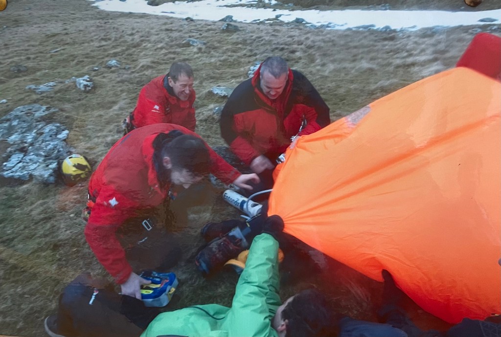
My group shelter came out of my rucksack and was deployed around me to protect me from the environment.
That shelter was not simply about comfort.
It was March in North Wales.
Two days earlier it had been snowing.
There was still snow on the surrounding ground.
It was around three o’clock in the afternoon and daylight would not last indefinitely.
Once you are immobilised on the ground, the thermal equation changes very quickly.
You are no longer generating meaningful heat by walking.
Conduction, convection, wind and exposure begin working against you.
I was getting colder.
I was actively shivering, partly because of the environment and partly because of the physiological response to trauma.
Using the JOMO Solutions Snowdonia mapping app on my phone, I established our exact grid reference. That information was relayed to Mountain Rescue and used to help determine how I was going to be extracted.
It would take approximately two hours for help to reach me.
So we waited.
The others kept me awake and checked my pulse and respirations roughly every ten minutes.
Meanwhile, I made three telephone calls.
The first was to my mother in Birmingham.
The second was to my father-in-law in London, because somebody was going to have to take my children to school for the foreseeable future.
The third was to my boss.
There seemed very little prospect of my arriving at work on Monday morning.
There is something quite surreal about lying on a cold Welsh hillside with a tibia and fibula in pieces while simultaneously organising the school run and reporting sick for work.
Eventually, we heard the helicopter.
“I Expect Nothing Less Than a Green”
When the medic was lowered down to me, something else happened almost automatically.
I stopped being quite so much the casualty and slipped back into professional mode.
“Hello Richard. I’m going to need to put a small blue cannula in you.”
“Blue cannula?”
I looked at my arms.
“Look at these veins. They’re like tree trunks. I expect nothing less than a green.”
Twenty years earlier, a consultant had taught me one of those dreadful little theatre mnemonics for remembering cannula sizes:
Blue is for boys.
Pink is pathetic.
Green is good.
Grey is great.
Orange is orgasmic.
Green it was.
And remarkably, that cannula remained in place throughout the entire clinical episode.
Then came the destruction of my expensive mountaineering equipment.
“I’m sorry, Rich. I’ve just cut off your gaiter.”
“You bastard. They’re Velcroed up the front!”
They were Gore-Tex Mountain Hardwear gaiters costing around £150.
A short pause.
“It’s worse than that, Rich. I’ve just cut off your socks.”
Smartwool Mountaineer socks.
About £30 a pair.
Only a week old.
At that particular moment, I was apparently still sufficiently compos mentis to mourn my socks.
Then came intravenous morphine and ketamine.
After that, I cared considerably less.
An inflatable splint was applied and I was prepared for extraction.
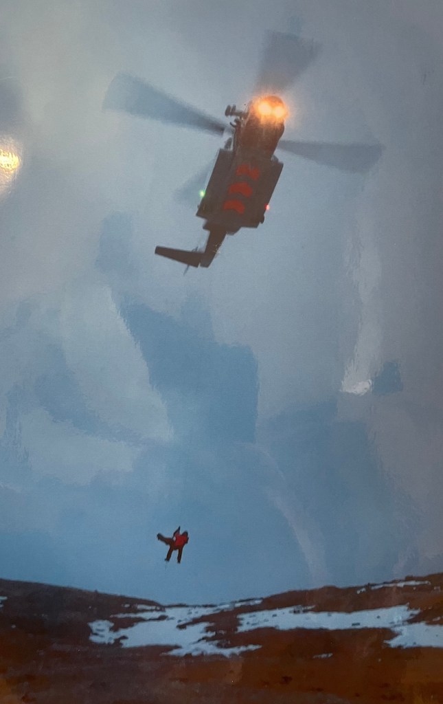
I was winched from the hillside into the helicopter.
Once aboard, I found myself sitting on the floor with what amounted to a Gore-Tex duvet wrapped around my shoulders.
After several hours of cold, it felt glorious.
Then I looked down at my leg.
My lower leg and foot appeared to be sitting at roughly ninety degrees of external rotation relative to my knee.
And instead of being horrified, I found it fascinating.
That is one of the extraordinary characteristics of ketamine in trauma medicine.
There can be profound analgesia accompanied by a dissociative quality that alters your emotional relationship with what is happening.
I knew it was my leg.
I knew exactly how serious the injury was.
But I could look at it almost academically.
I do not remember the flight.
The next clear memory I have is looking at the medic and saying:
“We’re at the hospital.”
“Rich, we’ve been here for thirty minutes.”
Bangor
Accident and Emergency was overwhelmed.
I was transferred onto a trolley and taken into a corridor area close to X-ray.
What I did not know was that my mother had driven from Birmingham to Bangor and had arrived perhaps ten minutes before the helicopter.
She had already primed the hospital staff.
Her summary was apparently something along the lines of:
Band 7 Operating Department Practitioner.
Morphine and ketamine.
Expect to be insulted.
A very pleasant young woman came to assess me.
I asked to see her ID badge.
“Ah. ST5 Orthopaedics.”
I looked at her.
“You might actually know something.”
Fortunately, she laughed.
We discussed the options.
One was immobilisation in a long-leg cast, effectively hip to foot, followed by transfer back towards London for definitive management.
The other was intramedullary fixation.
“Nail, please. Where do I sign?”
Consent signed.
Listed for the following day.
And suddenly an operation I understood intimately became entirely different.
I was no longer the ODP.
My name was on the theatre list.
The Anaesthetic I Had Prepared for Other People
The anaesthetic side was completely familiar to me.
I had prepared countless anaesthetic rooms.
I had checked machines.
Airway equipment.
Suction.
Monitoring.
Cannulas.
Drugs.
Emergency equipment.
I had stood beside countless patients while they surrendered consciousness to someone else.
Normally, I would be the person helping prepare the environment in which that surrender of control took place.
This time, I was lying on the trolley.
In my case, the anaesthetic was a general anaesthetic using a laryngeal mask airway and total intravenous anaesthesia: LMA and TIVA.
From a professional perspective, there was nothing exotic about that.
An LMA provides a supraglottic airway without the need for tracheal intubation in an appropriately selected patient.
TIVA means anaesthesia is maintained intravenously rather than predominantly with volatile inhalational agents.
From the ODP side, these are familiar techniques.
You understand the equipment.
You understand the drugs.
You understand the monitoring.
You know what the anaesthetist is trying to achieve and what the anaesthetic practitioner will be anticipating.
From the patient side, something much more fundamental is happening.
However experienced you are, you are surrendering control.
Your airway.
Your ventilation.
Your circulation.
Your consciousness.
Your safety.
I knew what every monitor was doing.
I knew what the airway device was for.
I knew what intravenous anaesthesia meant.
I knew what was likely being checked around me.
And then the drugs went in.
I was unconscious.
Professional knowledge does not remove that surrender of control.
If anything, knowing precisely what is happening makes the moment rather more interesting.
For years I had watched patients disappear under anaesthesia.
Now it was my turn.
A Tibial Nail From the Scrub Side
Once anaesthetised, the operation moved into territory I knew just as well from another part of my professional life.
From the perspective of an experienced orthopaedic scrub practitioner, tibial nailing has a familiar rhythm.
Position.
Image intensifier.
Entry point.
Guidewire.
Reduction.
Measure.
Ream.
Nail.
Lock.
Image.
Close.
That apparent simplicity hides a considerable amount of technical detail.
The patient is generally positioned supine on a radiolucent operating table with sufficient freedom for repeated movement of the image intensifier.
The proximal tibial entry point is identified under fluoroscopic guidance.
The cortex is opened.
A guidewire is introduced into the medullary canal.
For a reamed nail, a ball-tipped guidewire is passed through the reduced fracture and positioned within the distal tibia.
From the scrub side, this is the point at which one becomes obsessively protective of a piece of wire.
Lose the guidewire and everybody knows about it.
Once reduction and guidewire position are satisfactory, the appropriate nail length is determined.
Then comes reaming.
A flexible cannulated reamer is passed over the guidewire and the medullary canal is progressively enlarged.
Eight.
Eight and a half.
Nine.
Nine and a half.
Ten.
Ten and a half.
And eventually that familiar word:
“Chatter.”
An experienced scrub practitioner does not simply pass instruments when requested.
You know which reamer has just been used.
You know which comes next.
You watch the guidewire.
You watch the flutes.
You recognise when resistance feels wrong.
You already have a fairly good idea what diameter nail is likely to be selected.
Once the canal has been appropriately prepared, the selected tibial nail is assembled onto the insertion handle and targeting jig.
At this point the scrub practitioner should know exactly what is being implanted.
Correct system.
Correct nail.
Correct length.
Correct diameter.
Correct locking options.
Correct instrumentation.
The nail is introduced over the guidewire and advanced down the tibial canal while reduction is maintained.
Sometimes it advances relatively easily.
Sometimes it requires persuasion.
And orthopaedic persuasion frequently involves a hammer.
And, at times, an astonishing amount of brute force.
That is one of the things patients rarely appreciate about major orthopaedic surgery.
Under controlled anaesthesia, the human body can tolerate an extraordinary degree of manipulation: traction, rotation, reaming, drilling, hammering and repeated reduction manoeuvres that would be completely intolerable in an awake patient.
Anaesthesia does far more than simply make the patient unconscious.
It suppresses the pain and physiological stress response that major surgical stimulation would otherwise provoke.
Without adequate anaesthesia and analgesia, severe nociceptive stimulation can produce marked sympathetic activation: tachycardia, hypertension, increased myocardial workload and potentially significant cardiovascular instability, particularly in a patient who is already traumatised or physiologically compromised.
From behind the drapes, though, it can still be extraordinary to watch.
A fractured limb may be pulled, rotated, reduced and reamed.
A substantial metal implant can then be driven down the medullary canal with a hammer.
At one end of the table, the surgical team is applying force.
At the other, the anaesthetic team is quietly maintaining oxygenation, ventilation, circulation, analgesia and unconsciousness.
One side can look brutally physical.
The other appears measured, controlled and calm.
Together they make it possible for the patient to tolerate something that would otherwise be unbearable and physiologically dangerous.
It is controlled violence made possible by controlled physiology.
Once seated, the nail is locked.
Proximal locking is generally performed using the targeting jig.
The sequence becomes almost automatic:
Sleeve.
Trocar.
Drill.
Depth gauge.
Screw.
Screwdriver.
Distal locking often involves freehand fluoroscopic technique.
The radiographer produces the perfect-circle image.
The drill is aligned through the circle.
Near cortex.
Nail.
Far cortex.
Measure.
Screw.
Image.
Repeat.
If the fracture remains distracted, the construct can be manipulated to restore better fracture apposition.
Then eventually come the final images.
Alignment satisfactory.
Nail satisfactory.
Locking screws satisfactory.
Fracture reduced.
Wounds irrigated.
Closure.
Dressings.
Counts complete.
Implants documented.
Patient to recovery.
For the scrub practitioner, that feels like the end of the case.
For the patient, it is almost the beginning.
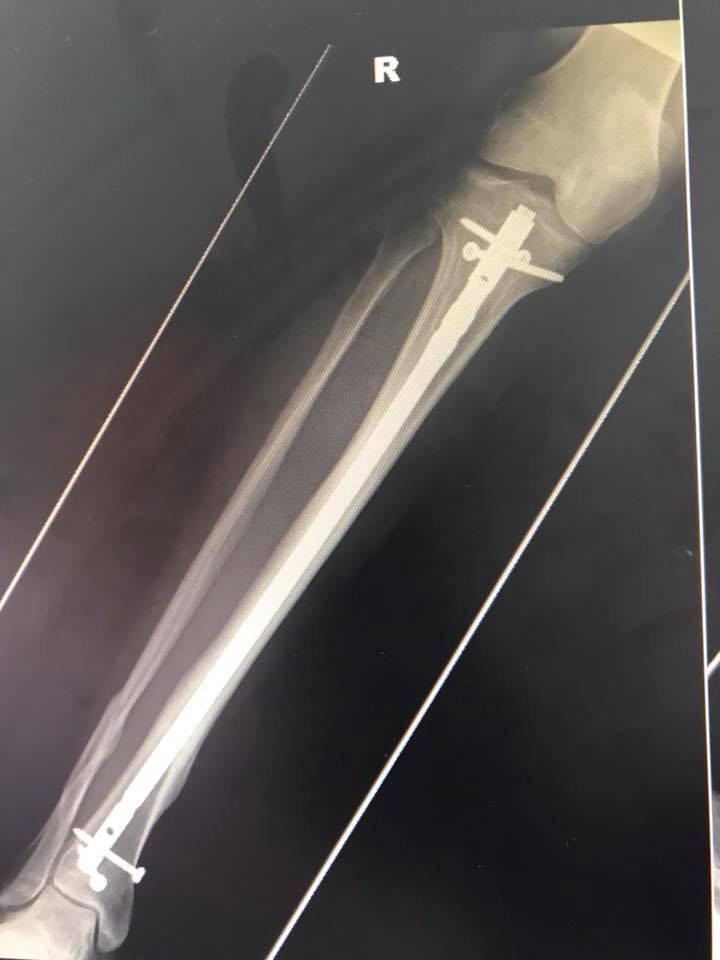
The Postoperative X-Ray
Eventually there was a postoperative radiograph.
I had looked at films like it countless times before.
A long intramedullary nail running down the tibia.
Proximal locking screws.
Distal locking screws.
Fracture spanned.
Alignment restored.
Normally, I would look at an image like that technically.
Reduction.
Nail position.
Screw position.
Length.
Alignment.
This time I was looking at the inside of my own leg.
The implant was no longer something whose batch number went onto a sticker.
It was now part of me.
And there is something else you understand rather differently once you become the patient.
Orthopaedic surgery is controlled trauma used to treat uncontrolled trauma.
The medullary canal has been entered.
The tibia has been reamed.
A metal nail has been advanced down its length.
Transverse screws have been drilled through bone and implant.
The fracture has been manipulated.
The limb has undergone both the original injury and the controlled additional trauma required to repair it.
When patients tell us that orthopaedic surgery hurts, perhaps we should occasionally remember precisely what we have done to them.
“A Long Time”
Postoperatively, the consultant seemed very keen to move me through the system.
I asked him a straightforward question.
“How long before I can return to work?”
His response was not particularly impressive.
“A long time.”
“What’s that supposed to mean? I’ve asked you a question. I expect a realistic answer.”
“Maybe six months.”
End of conversation.
As soon as physiotherapy had demonstrated that I could get myself up and down a flight of stairs on crutches, my bed was apparently required for the next hip fracture.
I was discharged on oxycodone and transferred into the nominal care of my local London hospital.
There was no structured rehabilitation programme.
No meaningful physiotherapy package.
No carefully planned return to activity.
No follow-up from Bangor.
I went home.
Fortunately, I could stay with my mother in Birmingham for a week before she drove me back down to London.
But I had already been given plenty of information about what I apparently would not be doing again.
I would not ski again.
I would not cycle again.
I would not mountaineer again.
I would not rock climb again.
I would be off work for at least six months.
The underlying message seemed to be:
You have broken your leg badly.
Sit down.
Accept it.
Wait.
That approach did not suit me.
Pain Is Weakness Leaving the Body!
Taken literally, the phrase is nonsense.
Pain is not simply weakness leaving the body.
Pain is a physiological warning system.
Pain after trauma has meaning.
Pain after surgery has meaning.
Pain can reflect tissue damage, inflammation, swelling, mechanical stress or complication.
Ignoring pain blindly is not bravery.
It is stupidity.
But pain can become something rather different during rehabilitation.
It can become uncertainty.
Fear.
Loss of confidence.
The constant question:
“Am I damaging this, or am I simply asking it to work again?”
That distinction matters.
I bought a turbo trainer for my road bike.
That became my self-imposed physiotherapy.
Not because I believed I knew better than every physiotherapist or orthopaedic surgeon.
But because there effectively was no rehabilitation programme.
And I understood enough anatomy, physiology and orthopaedics to recognise that doing absolutely nothing was not going to restore the life I wanted.
Recovery became progressive.
Movement.
Range.
Loading.
Strength.
Endurance.
Confidence.
Then more.
There is an enormous difference between progressive rehabilitation and simply ignoring an injury.
A healing tibia has biological limits.
Pain matters.
Swelling matters.
Radiographic union matters.
Delayed union, non-union, malalignment, infection and hardware problems are genuine complications.
But prolonged unnecessary inactivity also has consequences.
Muscle wasting.
Cardiovascular deconditioning.
Loss of proprioception.
Joint stiffness.
Fear of movement.
Loss of confidence.
A patient may eventually become radiographically healed while remaining functionally diminished.
That was not the outcome I wanted.
Four Months
Four months after the fracture, I returned to work.
I returned to cycling.
Clipped into the pedals.
Thirty miles a day.
At around nine months I was making a night descent of Glyder Fach.
The following summer I spent a week completing Via Ferrata and Klettersteig routes in the Bernese Oberland.
The following February I was skiing in Italy.
And I returned to climbing and mountaineering.
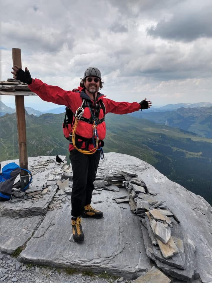
Eventually I stood on the summit of the Eiger Rotstock.
Helmet on.
Harness on.
Mountain boots on.
Arms spread wide.
That photograph means considerably more to me than any postoperative X-ray.
Because it demonstrates something an X-ray cannot.
It demonstrates that I got my life back.
What Does Successful Treatment Actually Mean?
None of this means every patient with a tibial fracture should ignore medical advice, buy a turbo trainer and head for the Alps.
That would be ridiculous.
Patients differ.
Fractures differ.
Healing rates differ.
Comorbidities differ.
Complications differ.
What my experience changed was my understanding of what we actually mean by a successful outcome.
In theatre, success can look like this:
Good reduction.
Correct nail.
Good alignment.
Locking screws satisfactory.
Wounds closed.
Patient stable.
Case finished.
Those outcomes matter enormously.
But they are not necessarily the outcomes that matter most to the person lying on the table.
For one patient, success may mean walking independently to the shops.
For another, returning to employment.
For another, playing with their grandchildren.
For an athlete, it may mean returning to competition.
For me, it meant cycling, skiing, climbing and mountaineering.
The patient’s pre-injury life should therefore form part of the rehabilitation conversation.
There is an enormous difference between saying:
“You will never do that again.”
and saying:
“This was a serious injury. I cannot promise how far you will recover, but we will rehabilitate you progressively and see what you can safely achieve.”
The first statement closes a door.
The second provides a pathway.
What Becoming the Patient Taught the ODP
Before my accident, I understood tibial nailing.
Technically, I probably understood considerably more about it than most patients ever would.
I understood the anaesthetic.
I understood the scrub procedure.
I understood the instruments.
I understood the implant.
I understood reaming.
I understood distal locking.
I understood the postoperative X-ray.
What I had not understood was the experience.
I had never heard my own bones snap.
I had never lain helpless on a cold hillside knowing precisely what had happened inside my own leg.
I had never looked at my own grossly rotated lower limb under ketamine and found myself clinically fascinated by it.
I had never handed control of my airway and consciousness to an anaesthetic team while knowing exactly what they were about to do.
I had never contemplated a reamer passing down my own tibia.
I had never looked at an intramedullary nail on an X-ray and realised that the implant I had handled countless times was now inside me.
And I had never properly appreciated the difference between fixing a fracture and restoring a person.
The intramedullary nail gave my tibia mechanical stability.
The surgical team did its job.
But getting my life back happened afterwards.
On crutches.
On stairs.
On a turbo trainer.
On a bicycle.
On wet Welsh ground.
On Via Ferrata cables.
On skis.
And eventually, once again, on mountain summits.
Perhaps “pain is weakness leaving the body” is nonsense.
Perhaps the more accurate version is this:
Pain tells you something has happened.
Recovery determines what happens next.
The surgeon’s postoperative X-ray tells us whether the operation has worked.
It does not necessarily tell us whether the treatment has succeeded.
Sometimes success is radiographic union.
Sometimes it is returning to work.
Sometimes it is clipping back into a bicycle pedal.
And sometimes it is standing on the summit of the Eiger Rotstock with a tibial nail inside your leg, looking across the Alps and realising that the fracture never got to decide who you were.