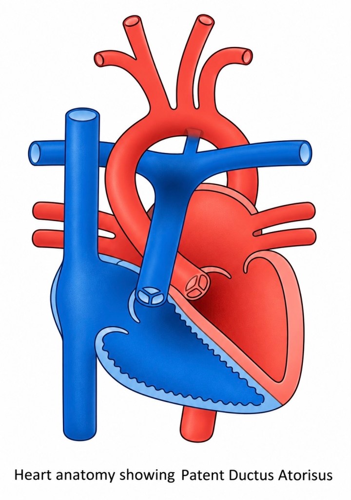
A Patent Ductus Arteriosus (PDA) occurs when the ductus arteriosus remains patent after birth rather than undergoing the normal process of closure.
The significance of a PDA depends upon its size, the age of the patient, pulmonary vascular resistance and the pressure difference between the systemic and pulmonary circulations.
Immediately after birth, the direction of ductal flow changes.
Because aortic pressure is normally greater than pulmonary arterial pressure, blood flows from the aorta through the PDA into the pulmonary artery.
This creates a left-to-right shunt.
In simple terms, oxygenated blood that has already passed through the lungs is sent back towards the lungs again.
A small PDA may produce relatively little haemodynamic disturbance.
A large PDA, however, can result in substantial pulmonary over-circulation.
More blood returns through the pulmonary veins to the left atrium and left ventricle, increasing left-sided volume loading.
Over time, persistently excessive pulmonary blood flow can contribute to pulmonary vascular remodelling and pulmonary hypertension.
⸻
PDA and Eisenmenger Physiology
If a large left-to-right shunt remains untreated for a sufficiently long period, pulmonary vascular resistance can progressively rise.
Eventually pulmonary arterial pressure may approach or exceed systemic pressure.
The original left-to-right shunt can consequently become bidirectional and ultimately reverse.
Blood then passes from the pulmonary artery into the systemic circulation.
This is a right-to-left shunt.
Deoxygenated blood consequently enters the systemic circulation and cyanosis develops.
This process forms part of Eisenmenger syndrome.
The important correction to some older descriptions is that Eisenmenger syndrome is associated with pulmonary hypertension, not pulmonary hypotension.
Once severe irreversible pulmonary vascular disease has developed, simply closing the original communication can be dangerous because the defect may effectively have become a pressure-relief pathway for the right side of the circulation.
⸻
Closure of a PDA
The appropriate treatment depends heavily upon gestation, age, anatomy and haemodynamic significance.
In premature neonates, pharmacological treatment may be used to encourage ductal closure. Drugs that inhibit prostaglandin synthesis can promote constriction of the ductus.
In other circumstances, PDA closure may be achieved using a catheter-delivered occlusion device.
Surgical ligation or division remains an option where catheter treatment is unsuitable or where the clinical circumstances require surgical management.
Historically, surgical PDA ligation was performed through a left posterolateral thoracotomy without cardiopulmonary bypass.
The ductus is identified between the pulmonary artery and descending aorta before being clipped, ligated or divided.
The key physiological principle, however, is considerably simpler than the operation:
Before birth, the ductus is necessary. After birth, continued patency may create an abnormal pathway between the high-pressure systemic circulation and the lower-pressure pulmonary circulation.
⸻
Atrial Septal Defect — ASD
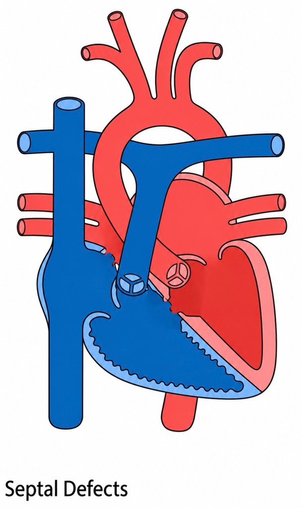
An Atrial Septal Defect (ASD) is an abnormal communication between the right and left atria.
Following the normal postnatal fall in pulmonary vascular resistance, pressure within the left atrium generally exceeds pressure within the right atrium.
Blood therefore tends to pass through an ASD from left to right.
Again, oxygenated blood is effectively being recirculated through the pulmonary circulation.
Unlike a large VSD, the pressure difference between the atria is relatively small.
The importance of an ASD is therefore determined less by a large pressure gradient across the defect and more by the volume of blood being shunted.
A significant ASD produces increased volume loading of the right atrium and right ventricle and increased pulmonary blood flow. (Merck Manuals)
⸻
Types of ASD
ASDs are classified according to their anatomical position.
Ostium Secundum ASD
The secundum ASD is the most common type.
It occurs in the region of the fossa ovalis, approximately centrally within the atrial septum.
Many secundum defects can be suitable for transcatheter device closure if their size and surrounding septal margins are appropriate. (Merck Manuals)
Ostium Primum Defect
The ostium primum defect lies low in the atrial septum, adjacent to the atrioventricular valves.
It is important not to regard this simply as another variety of isolated ASD.
A primum defect belongs anatomically to the spectrum of atrioventricular septal defects (AVSD) and may be associated with abnormalities of the atrioventricular valves. (Merck Manuals)
Sinus Venosus Defect
A sinus venosus defect is usually located close to the entry of the superior vena cava into the right atrium, although inferior forms also occur.
These defects are frequently associated with anomalous pulmonary venous drainage, particularly of the right-sided pulmonary veins. (Merck Manuals)
This association is important surgically because correction may involve not merely closing a hole but redirecting pulmonary venous blood appropriately into the left atrium.
⸻
Clinical Effects of an ASD
Children with an ASD may remain asymptomatic for many years.
A significant defect nonetheless produces continued right-heart volume loading.
Over time this may result in right atrial and right ventricular enlargement, pulmonary hypertension and atrial arrhythmias.
Very longstanding large shunts may eventually lead to severe pulmonary vascular disease, although progression to Eisenmenger physiology is considerably less common with ASD than with large ventricular or great-vessel communications. (Merck Manuals)
Diagnosis is principally established by echocardiography, which identifies the location and size of the defect, the direction of the shunt and its effects upon the cardiac chambers.
⸻
ASD Closure
Not every ASD requires intervention.
Small haemodynamically insignificant communications can be observed, particularly in young children.
Where the defect produces significant right-heart volume overload, closure is normally considered.
Appropriately located secundum ASDs may be closed using a transcatheter occlusion device.
Defects that are unsuitable for device closure, including most primum and sinus venosus defects, require surgical repair. (Merck Manuals)
Surgical closure is normally performed on cardiopulmonary bypass.
Depending upon size and anatomy, the defect may be closed directly with sutures or with a patch, commonly using autologous pericardium or synthetic material.
Again, the central physiological idea is straightforward:
An ASD permits blood to move between the atria. After birth this is normally a left-to-right shunt, producing excessive pulmonary blood flow and volume loading of the right heart.
⸻
Ventricular Septal Defect — VSD
A Ventricular Septal Defect (VSD) is an abnormal communication between the right and left ventricles.
VSDs vary enormously in clinical importance.
A tiny muscular VSD may have little physiological consequence and may close spontaneously.
A large non-restrictive VSD can profoundly alter the circulation.
The key difference between an ASD and a VSD is the pressure environment in which the defect exists.
The ventricles generate considerably greater pressures than the atria.
After pulmonary vascular resistance falls following birth, left ventricular pressure is normally substantially higher than right ventricular pressure.
Blood therefore travels through a VSD from the left ventricle into the right ventricle.
This creates a left-to-right ventricular shunt.
⸻
Size Versus Physiological Size
The physical diameter of a VSD is important, but its physiological effect is determined by more than measurement alone.
A small defect may restrict blood flow and maintain a considerable pressure difference between the ventricles.
This is described as a restrictive VSD.
A very large defect may allow ventricular pressures to approach equalisation.
This is described as a non-restrictive VSD.
Consequently, a small VSD may produce a loud murmur because blood passes through it at high velocity while producing relatively little overall haemodynamic disturbance.
Conversely, a very large VSD can be physiologically dangerous despite sometimes generating less impressive turbulence.
⸻
Types of VSD
VSDs are described according to their anatomical position within the ventricular septum.
Common categories include:
Perimembranous VSDs
Located in the membranous region of the septum and among the most commonly encountered forms.
Muscular VSDs
Located entirely within muscular septal tissue. They may occur in different regions and may occasionally be multiple, producing the so-called “Swiss cheese” septum.
Inlet VSDs
Located posteriorly beneath the atrioventricular valves and associated particularly with the AVSD spectrum.
Outlet or subarterial VSDs
Located close to the ventricular outflow tracts and semilunar valves.
The anatomical location matters not simply for classification but because it influences associated abnormalities, likelihood of spontaneous closure, suitability for device closure and the surgical approach required.
⸻
Haemodynamic Consequences of a Large VSD
With a large VSD, substantial quantities of blood may travel from the left ventricle into the right ventricle and subsequently into the pulmonary circulation.
This results in pulmonary over-circulation.
The increased pulmonary venous return then reaches the left atrium and left ventricle, producing left-sided volume loading.
Infants with a significant VSD may therefore develop symptoms of heart failure as pulmonary vascular resistance falls during the first weeks of life.
Typical features can include:
- tachypnoea;
- increased work of breathing;
- sweating during feeding;
- prolonged or tiring feeds;
- poor weight gain;
- and failure to thrive.
Large untreated VSDs can expose the pulmonary vascular bed to both excessive flow and pressure.
Over time this can cause pulmonary vascular remodelling and progressive pulmonary hypertension.
If pulmonary vascular resistance eventually exceeds systemic vascular resistance, the shunt may reverse from right to left and produce Eisenmenger physiology.
⸻
Spontaneous Closure
Many small VSDs, particularly muscular defects, become smaller or close spontaneously during childhood.
The likelihood of spontaneous closure depends upon the location and anatomy of the defect.
A small asymptomatic VSD with no adverse haemodynamic effects may therefore require observation rather than intervention.
Large defects producing significant pulmonary over-circulation, heart failure, failure to thrive or rising pulmonary pressures are managed very differently.
⸻
Closure of a VSD
Surgical closure remains an important treatment for significant VSDs.
The heart is supported with cardiopulmonary bypass while the surgeon approaches the defect, commonly through the right atrium and tricuspid valve where anatomy permits.
A patch is then secured around the margins of the defect.
The position of the cardiac conduction system is particularly important because part of the conduction pathway runs close to the margins of certain perimembranous VSDs.
Damage can produce atrioventricular block.
Some selected VSDs may also be suitable for transcatheter device closure, depending upon their anatomy and location.
The central principle remains:
A VSD provides a communication between two powerful pumping chambers. Its consequences depend upon the size of that communication and, crucially, upon the pressure and resistance on either side of it.
⸻
Bringing the Physiology Together
PDA, ASD and VSD appear anatomically very different.
Physiologically, however, they share an important concept.
Each creates an abnormal communication between two parts of the circulation.
After birth the normal relationship is:
Systemic vascular resistance > pulmonary vascular resistance.
Consequently, blood passing through many uncomplicated congenital communications initially travels from the systemic or left side of the circulation towards the pulmonary or right side.
This produces a left-to-right shunt.
The patient may therefore have substantial congenital heart disease without initially appearing cyanosed.
The problem is not necessarily inadequate oxygenation of the blood.
The problem may instead be that too much blood is repeatedly circulating through the lungs.
If excessive pulmonary blood flow and pressure continue for long enough, the pulmonary vascular bed can progressively remodel.
Pulmonary vascular resistance rises.
Eventually the original direction of the pressure gradient can disappear or reverse.
A right-to-left shunt then allows deoxygenated blood into the systemic circulation.
Only at this point may a previously acyanotic defect become cyanotic.
Understanding this progression is considerably more useful than simply memorising lists of cyanotic and acyanotic congenital heart defects.
It returns congenital cardiac physiology to its most fundamental principles:
Pressure. Resistance. Direction of flow. Oxygenation.
If those four things can be established, much of what initially appears extraordinarily complicated begins to make sense.
⸻
Glossary of Common Terms
Aortopulmonary Window — AP Window
An abnormal communication between the ascending aorta and pulmonary artery.
⸻
Acyanotic
A congenital cardiac condition in which systemic arterial oxygenation is generally maintained and the patient does not characteristically demonstrate central cyanosis.
The term does not mean excessive oxygenation.
⸻
Atresia
Congenital absence or failure of normal formation of an opening, valve or vascular connection.
⸻
Atrial Septal Defect — ASD
An abnormal communication between the right and left atria.
⸻
Atrioventricular Septal Defect — AVSD
A group of congenital defects involving deficient formation of the atrioventricular septal structures and abnormalities of the atrioventricular valve junction.
AVSD may be partial, transitional or complete.
⸻
Blalock–Taussig–Thomas Shunt — BTT Shunt
A systemic-to-pulmonary arterial shunt designed to increase pulmonary blood flow.
Modern forms usually use a synthetic graft between a systemic artery and a pulmonary artery rather than directly joining the subclavian artery to the pulmonary artery.
⸻
Cardiopulmonary Bypass — CPB
A technique in which a heart-lung machine temporarily maintains circulation and gas exchange while cardiac surgery is undertaken.
⸻
Coarctation of the Aorta — CoA
Congenital narrowing of the aorta, typically in the region of the aortic isthmus.
⸻
Cyanosis
Bluish discoloration of the skin or mucous membranes resulting from increased quantities of deoxygenated haemoglobin within the circulation.
⸻
Ductus Arteriosus
A normal foetal vascular connection between the pulmonary artery and descending aorta.
⸻
Eisenmenger Syndrome
Severe pulmonary vascular disease resulting from a longstanding systemic-to-pulmonary shunt, in which pulmonary vascular resistance rises sufficiently to produce bidirectional or reversed shunting and cyanosis.
⸻
Foramen Ovale — FO
A normal foetal communication between the right and left atria that allows blood to bypass the foetal pulmonary circulation.
⸻
Hypoplastic Left Heart Syndrome — HLHS
A spectrum of congenital abnormalities characterised by severe underdevelopment of left-sided cardiac structures, resulting in an inability of the left ventricle to support the systemic circulation adequately.
⸻
Interrupted Aortic Arch — IAA
Complete discontinuity between portions of the aortic arch.
⸻
Major Aortopulmonary Collateral Arteries — MAPCAs
Abnormal systemic arteries supplying pulmonary blood flow, most commonly encountered in association with complex congenital heart disease such as pulmonary atresia with VSD.
⸻
Mitral Atresia — MA
Failure of formation or complete obstruction of the normal mitral valve orifice.
⸻
Patent Ductus Arteriosus — PDA
Persistence of the ductus arteriosus after birth.
⸻
Pulmonary Atresia — PA
Absence of a patent connection through the pulmonary valve or right ventricular outflow, preventing normal forward blood flow from the right ventricle to the pulmonary artery.
⸻
Pulmonary Vascular Resistance — PVR
The resistance that the right ventricle must overcome to drive blood through the pulmonary circulation.
⸻
Shunt
Abnormal or deliberately created passage of blood from one part of the circulation to another.
The direction of shunting is determined primarily by pressure gradients and vascular resistance.
⸻
Stenosis
Abnormal narrowing of a valve, vessel or opening.
⸻
Systemic Vascular Resistance — SVR
The resistance presented by the systemic circulation to blood ejected by the left ventricle.
⸻
Tetralogy of Fallot — TOF
A congenital cardiac condition characterised by ventricular septal defect, overriding aorta, right ventricular outflow tract obstruction and right ventricular hypertrophy.
⸻
Total Anomalous Pulmonary Venous Return — TAPVR
A congenital abnormality in which all pulmonary veins return to the systemic venous circulation rather than connecting normally to the left atrium.
⸻
Transposition of the Great Arteries — TGA
A congenital abnormality in which the aorta arises from the right ventricle and the pulmonary artery arises from the left ventricle, producing two parallel circulations unless mixing occurs between them.
⸻
Tricuspid Atresia — TA
Congenital absence of a functional connection between the right atrium and right ventricle through the tricuspid valve.
⸻
Truncus Arteriosus
A congenital abnormality in which a single arterial trunk arises from the heart and supplies the systemic, pulmonary and coronary circulations because normal separation into the aorta and pulmonary artery has not occurred.
⸻
Ventricular Septal Defect — VSD
An abnormal communication between the right and left ventricles.
⸻
Source note
The physiological descriptions in this revised edition have been checked against contemporary descriptions of foetal/neonatal circulation and congenital septal defects. In particular, the revision corrects the original description of Eisenmenger syndrome as involving pulmonary “hypotension”; it is associated with progressive pulmonary hypertension and increased pulmonary vascular resistance. Current descriptions also distinguish true ASD from persistent or stretched foramen ovale and recognise ostium primum defects as part of the AVSD spectrum. (Merck Manuals)

