Richard Purchon FRGS AC RODP
Adult cardiac surgery encompasses a broad range of procedures designed to treat disease affecting the heart, its valves, the coronary circulation and the great vessels.
For those working within the cardiac operating theatre, understanding the operation requires more than simply knowing its name. It requires an appreciation of the anatomy, normal circulation, electrical activity of the heart, surgical approach, cardiopulmonary bypass, vascular conduits, valve pathology and the considerable physiological changes that occur when the heart is temporarily stopped, opened, repaired and restarted.
This article provides an introduction to some of the most commonly encountered areas of adult cardiac surgery, with particular emphasis on what members of the perioperative team are likely to see in theatre.
Normal Cardiac Anatomy
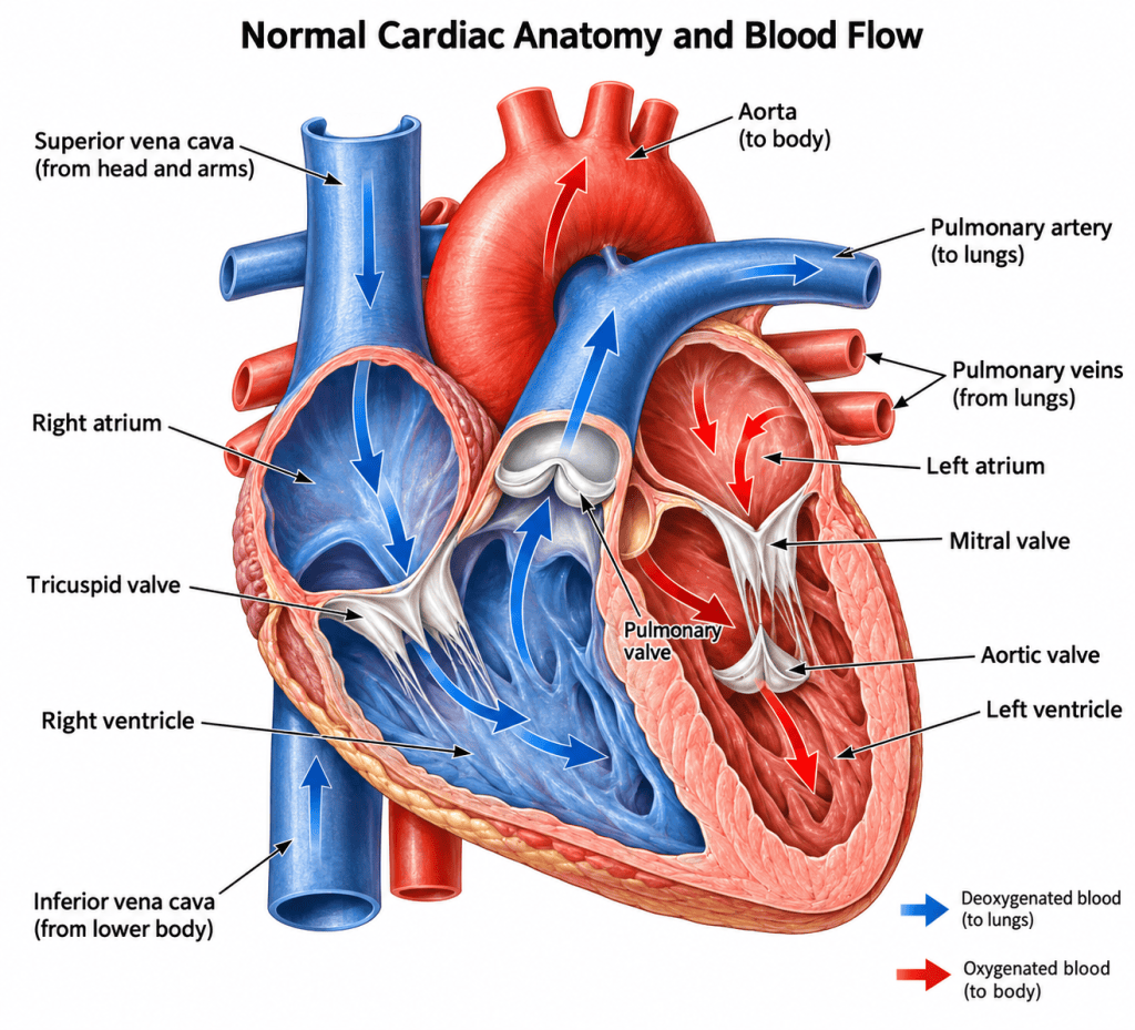
The heart is essentially two pumps working together.
The right side receives deoxygenated blood returning from the systemic circulation and pumps it through the pulmonary circulation to the lungs.
The left side receives oxygenated blood returning from the lungs and pumps it through the aorta to the systemic circulation.
Blood normally travels through the heart in the following sequence:
Superior and inferior vena cava → right atrium → tricuspid valve → right ventricle → pulmonary valve → pulmonary artery → lungs → pulmonary veins → left atrium → mitral valve → left ventricle → aortic valve → aorta
Four valves ensure that blood predominantly moves in one direction:
- Tricuspid valve
- Pulmonary valve
- Mitral valve
- Aortic valve
The right and left ventricles are anatomically and physiologically different.
The right ventricle pumps into the relatively low-pressure pulmonary circulation, whereas the muscular left ventricle must generate sufficient pressure to propel blood throughout the systemic circulation.
The Coronary Circulation
The myocardium requires its own blood supply.
The coronary arteries arise from the aortic root immediately above the aortic valve and distribute oxygenated blood throughout the myocardium.
The two principal coronary systems are the right coronary artery and left main coronary artery.
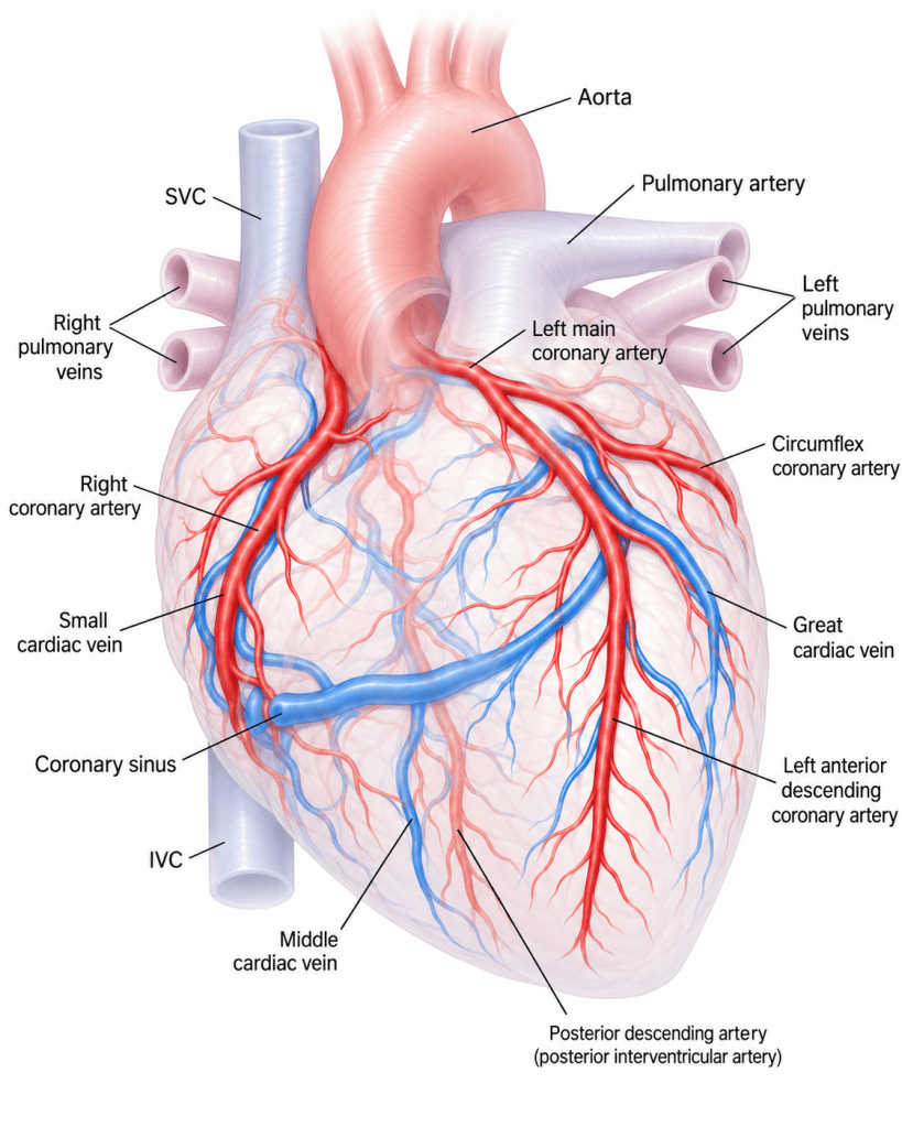
The left main coronary artery usually divides into:
- Left anterior descending artery – LAD
- Circumflex artery – CX
The LAD runs in the anterior interventricular groove and supplies a substantial proportion of the anterior left ventricle and interventricular septum.
The circumflex travels within the atrioventricular groove and supplies the lateral and, depending upon coronary dominance, portions of the posterior myocardium.
The right coronary artery travels within the right atrioventricular groove and normally supplies much of the right ventricle and inferior surface of the heart.
The exact distribution varies considerably between individuals.
Obstruction of these arteries through atherosclerotic coronary artery disease may reduce myocardial blood flow sufficiently to produce angina, myocardial infarction and impaired ventricular function.
Electrical Conduction of the Heart
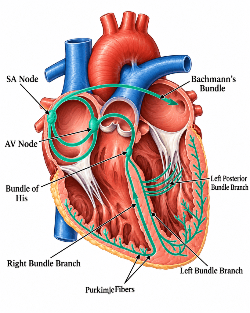
Mechanical contraction of the heart depends upon an organised electrical conduction system.
Under normal circumstances an impulse originates within the sinoatrial node – SA node, situated within the right atrium.
The impulse spreads through the atria, producing atrial depolarisation and contraction.
It subsequently reaches the atrioventricular node – AV node, where conduction slows briefly before passing through the:
Bundle of His → right and left bundle branches → Purkinje network
This rapid ventricular conduction allows coordinated ventricular depolarisation and contraction.
Relating Cardiac Conduction to the ECG
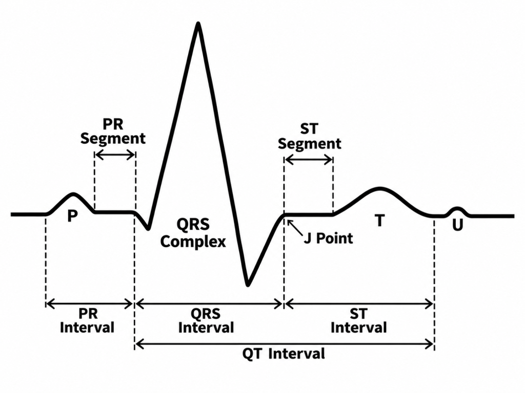
The electrocardiogram provides a surface representation of cardiac electrical activity.
The major components are:
P wave
Represents atrial depolarisation.
PR interval
Measured from the beginning of the P wave to the beginning of the QRS complex. A normal adult PR interval is approximately 0.12–0.20 seconds.
QRS complex
Represents ventricular depolarisation.
ST segment
Normally relatively isoelectric and represents the period following ventricular depolarisation before repolarisation becomes evident.
T wave
Represents ventricular repolarisation.
QT interval
Represents the overall period of ventricular depolarisation and repolarisation and varies significantly with heart rate.
Changes in rhythm, conduction, myocardial perfusion, electrolytes and temperature can all produce ECG abnormalities during cardiac surgery.
Surgical Access to the Heart
There is no single incision used for cardiac surgery.
The approach depends upon the structure requiring treatment, the operation being performed, previous surgery, the patient’s anatomy and the surgeon’s preferred technique.
Median Sternotomy
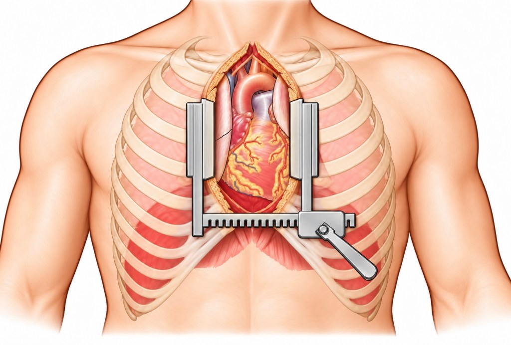
The traditional approach to cardiac surgery is the median sternotomy.
A midline incision is made over the sternum before the sternum itself is divided longitudinally.
A sternal retractor then separates the two halves, providing excellent exposure of:
- the heart
- ascending aorta
- right atrium
- venae cavae
- pulmonary artery
- coronary vessels
Median sternotomy remains particularly useful where extensive exposure is required, including:
- coronary artery bypass grafting
- multiple valve procedures
- aortic root and ascending aortic surgery
- complex redo procedures
- emergency surgery
Mini-Sternotomy
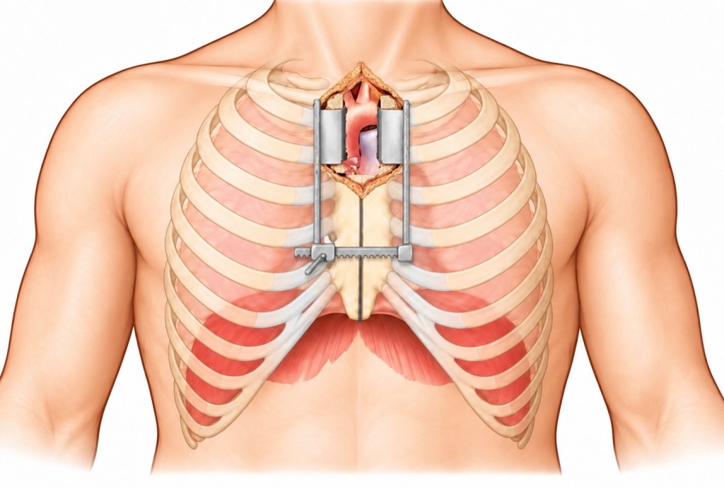
A mini-sternotomy provides access through only part of the sternum.
An upper partial sternotomy may be used for selected aortic valve and ascending aortic procedures.
The theoretical advantages of reduced-access approaches include:
- smaller incision
- reduced tissue disruption
- potentially reduced postoperative pain
- earlier mobilisation
- cosmetic benefits
The procedure is not simply a smaller version of conventional surgery. Restricted access changes instrument handling, cannulation strategy, surgical exposure and emergency planning.
Right Anterior Thoracotomy
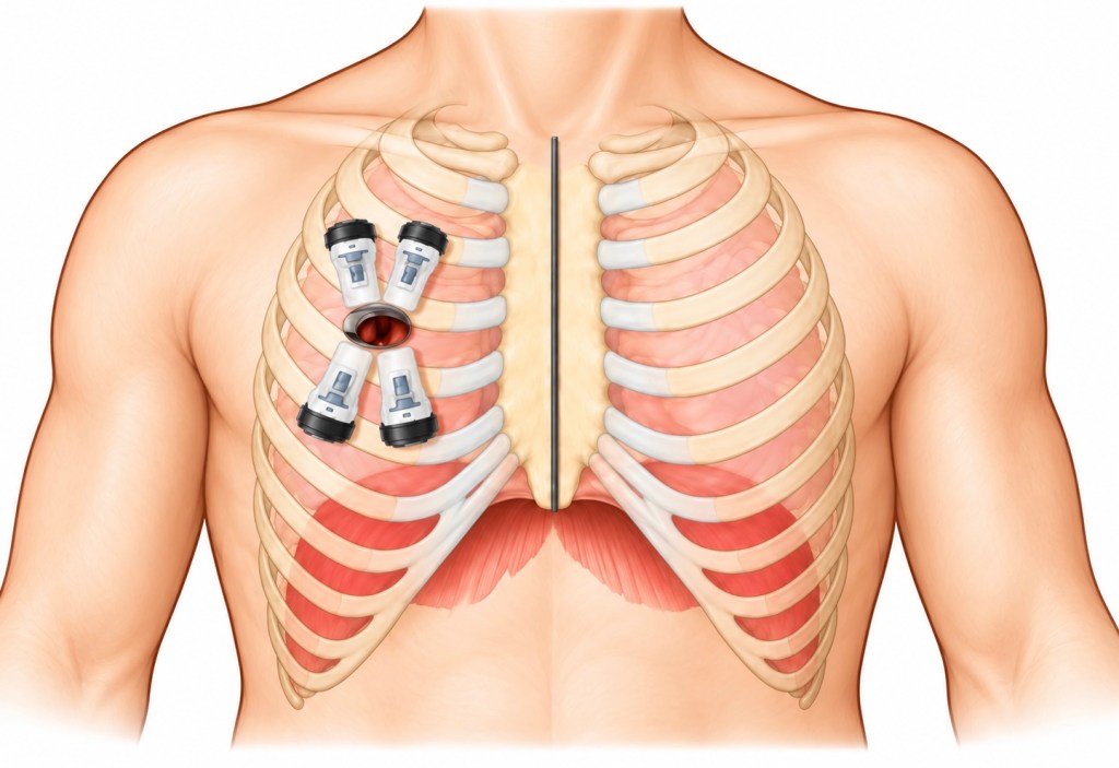
A right anterior thoracotomy provides another minimally invasive approach to the heart.
Access is obtained through an intercostal space rather than dividing the entire sternum.
It may be used for selected procedures, particularly minimally invasive aortic or mitral valve surgery depending upon the technique employed by the cardiac centre.
Peripheral cardiopulmonary bypass cannulation, frequently involving the femoral vessels, may be required.
Minimally Invasive Coronary Surgery
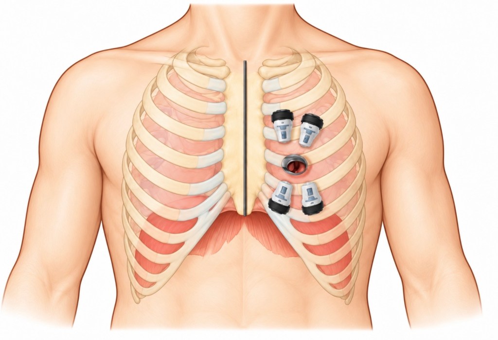
Selected coronary bypass procedures may also be performed through a limited thoracic incision.
MIDCAB – Minimally Invasive Direct Coronary Artery Bypass – is most commonly associated with bypass of the LAD using the internal mammary artery without conventional full sternotomy.
Endoscopic and robotic techniques can also be used to assist internal mammary artery harvesting.
These approaches require careful patient selection and specialist expertise.
Cardiopulmonary Bypass
Many cardiac operations require the surgeon to operate on a motionless and relatively bloodless heart.
This is made possible through cardiopulmonary bypass – CPB.
A heart-lung machine temporarily performs the principal circulatory functions normally provided by the heart and lungs.
Venous blood is diverted from the patient into the bypass circuit.
Within the circuit it is:
- collected
- oxygenated
- cleared of carbon dioxide
- temperature controlled
- pumped back into the arterial circulation
Cannulae are placed into appropriate venous and arterial structures.
Depending upon the operation, venous drainage may be obtained through the right atrium using a two-stage cannula or through separate superior and inferior vena caval cannulae.
Arterial return is commonly through the ascending aorta, although alternative cannulation sites may be required.
The Aortic Cross-Clamp and Cardioplegia
Once cardiopulmonary bypass has been established, many operations require the ascending aorta to be clamped.
The aortic cross-clamp separates the heart from the systemic circulation supplied by the bypass machine.
Because coronary blood flow would otherwise stop, the myocardium must be protected.
A cardioplegia solution is administered to intentionally arrest the heart and reduce myocardial metabolic demand.
Cardioplegia may be delivered:
- antegrade through the aortic root or coronary ostia
- retrograde through the coronary sinus
- or using a combination of techniques
It may be administered as blood cardioplegia, crystalloid cardioplegia or combinations depending upon institutional and surgical practice.
Myocardial protection is one of the fundamental components of safe cardiac surgery.
Coronary Artery Bypass Grafting
Coronary artery bypass grafting – CABG is performed to restore blood supply beyond significant obstructive coronary artery disease.
Rather than removing the coronary obstruction, the surgeon creates an alternative route for blood to reach the myocardium beyond the diseased segment.
Common conduits include:
- left internal mammary/internal thoracic artery – LIMA/LITA
- right internal mammary/internal thoracic artery – RIMA/RITA
- radial artery
- greater saphenous vein
The left internal mammary artery to the LAD is an important and widely used arterial graft.
Saphenous vein grafts are harvested from the leg, while the radial artery may be harvested from the forearm following appropriate assessment of the hand’s collateral circulation.
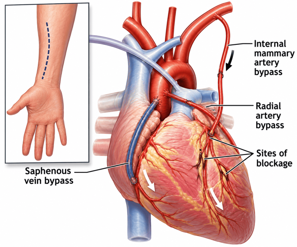
Proximal and Distal Anastomoses
A free bypass graft requires connections between the graft and the patient’s circulation.
The coronary connection beyond the obstruction is commonly referred to as the distal anastomosis.
For a saphenous vein or free arterial graft, the opposite end is normally attached to the aorta, forming the proximal anastomosis.
An internal mammary artery can remain attached to its natural origin and therefore usually requires only its distal end to be attached to the coronary artery.
Multiple distal anastomoses may sometimes be created from a single conduit in sequential grafting.
Valve Surgery
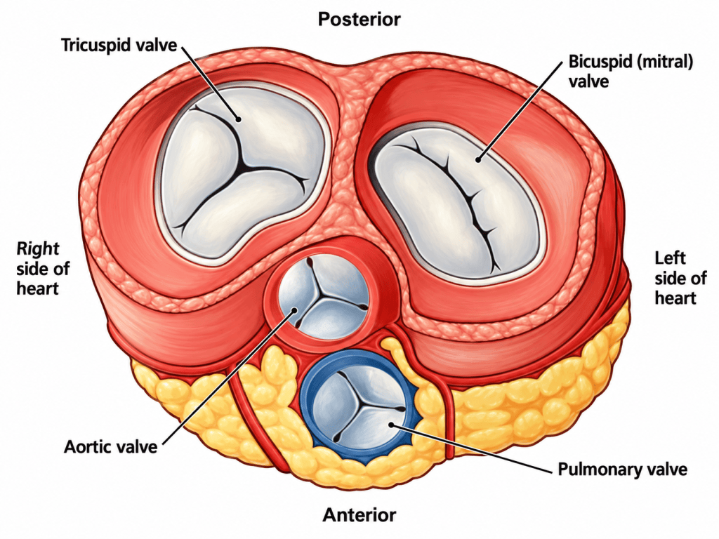
The heart contains four valves:
- aortic
- mitral
- tricuspid
- pulmonary
In adult cardiac surgery the aortic and mitral valves account for a large proportion of valve procedures, although tricuspid surgery is increasingly encountered.
Valve disease broadly produces two mechanical problems.
Stenosis
A stenotic valve does not open adequately.
The heart must therefore generate greater pressure to force blood through the narrowed opening.
For example, severe aortic stenosis increases left ventricular afterload and can eventually produce ventricular hypertrophy and cardiac failure.
Regurgitation
A regurgitant or incompetent valve fails to close effectively.
Blood therefore flows backwards across the valve.
The consequences depend upon the valve affected and the severity and duration of regurgitation.
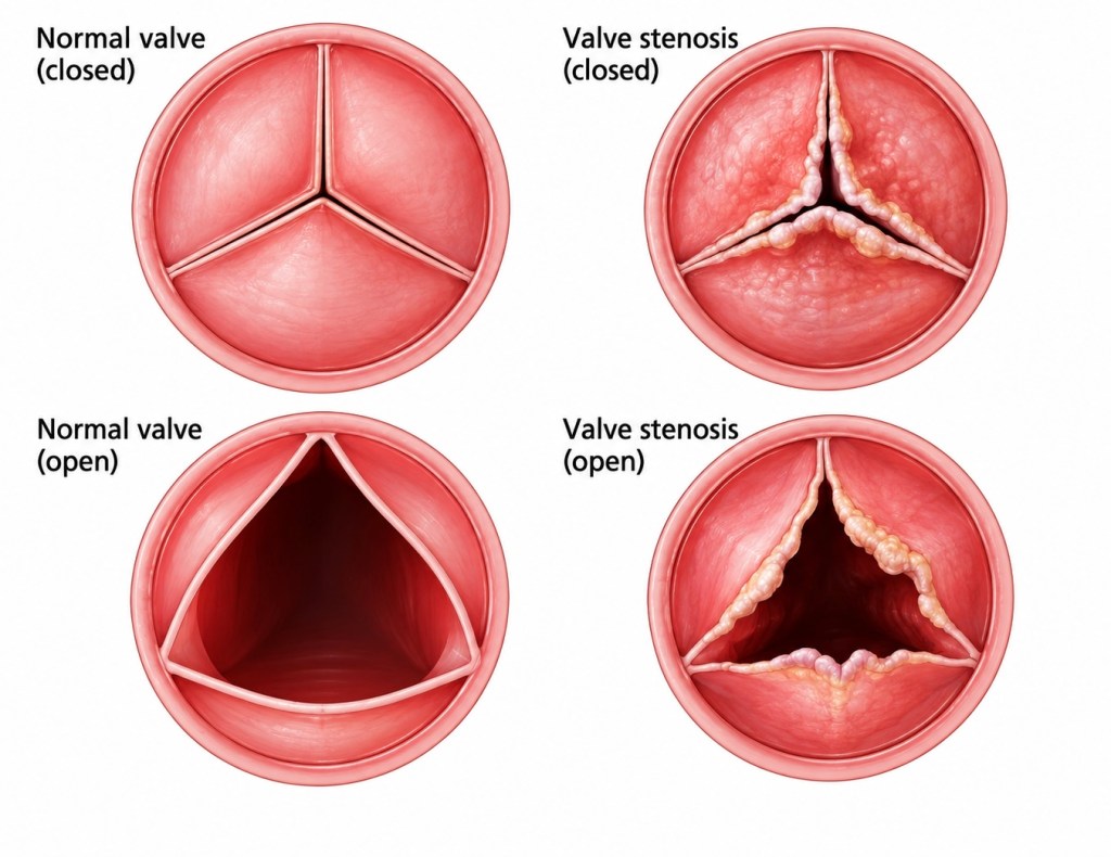
Comparison of normal and stenotic valve opening
Valve Repair or Replacement?
Whenever appropriate, surgeons may attempt to preserve and repair the native valve rather than replace it.
Mitral valve repair may involve:
- annuloplasty rings
- leaflet resection
- artificial chordae
- reconstruction of leaflet tissue
If repair is not possible or appropriate, valve replacement may be required.
Replacement valves broadly fall into two groups.
Mechanical Valves
Mechanical prostheses are extremely durable but generally require long-term anticoagulation because of their thrombogenic potential.
Biological Valves
Biological or tissue valves are constructed using biological material, commonly bovine pericardial or porcine tissue.
They usually require less intensive long-term anticoagulation specifically for the prosthesis but have limited structural durability compared with mechanical valves.
Valve choice therefore involves balancing age, durability, bleeding risk, anticoagulation, pregnancy considerations, patient preference and other clinical factors.
Transcatheter Aortic Valve Implantation
Not all valve replacements require open cardiac surgery.
TAVI – Transcatheter Aortic Valve Implantation, also commonly termed TAVR – Transcatheter Aortic Valve Replacement, allows a prosthetic aortic valve to be positioned using a catheter-based technique.
The transfemoral approach is commonly used, although alternative access routes may be required.
TAVI was originally developed predominantly for patients considered at prohibitive or very high surgical risk. Its use has subsequently expanded considerably, with patient selection now depending upon factors including age, anatomy, surgical risk, durability considerations and multidisciplinary Heart Team assessment.
Aortic Disease
Cardiac surgery also encompasses disease of the ascending aorta and aortic root.
This includes:
- aortic aneurysm
- acute aortic dissection
- aortic root dilatation
- connective-tissue disorders
- infection
- traumatic and degenerative disease
The aorta is exposed continuously to pulsatile arterial pressure.
Disease affecting the integrity of its wall can therefore have catastrophic consequences.
Aortic Dissection
An aortic dissection occurs when blood gains access to the layers of the aortic wall, producing a false lumen that can propagate along the vessel.
This may compromise:
- coronary arteries
- cerebral circulation
- spinal circulation
- renal arteries
- mesenteric vessels
- limb perfusion
It may also result in:
- acute aortic regurgitation
- cardiac tamponade
- rupture
- catastrophic haemorrhage
Two traditional classification systems are frequently encountered.
Stanford Classification
Type A
Any dissection involving the ascending aorta.
Type B
Dissection that does not involve the ascending aorta.
DeBakey Classification
Type I begins in the ascending aorta and extends beyond it.
Type II is confined predominantly to the ascending aorta.
Type III originates in the descending thoracic aorta and extends distally to varying degrees.
Acute Stanford Type A dissection is generally a surgical emergency.
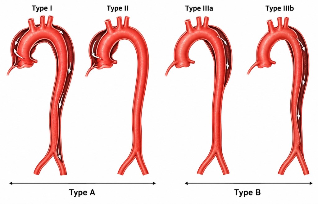
Stanford and DeBakey classification of Aortic Dissection
Anaesthetic Considerations in Acute Aortic Dissection
These patients can deteriorate extraordinarily quickly.
Depending upon the pathology and local practice, preparation may include:
- large-bore venous access
- invasive arterial monitoring
- consideration of bilateral arterial pressure monitoring
- central venous access
- transoesophageal echocardiography
- external defibrillation pads
- cerebral oxygenation monitoring where appropriate
- blood products and rapid-transfusion capability immediately available
- cardiopulmonary bypass equipment prepared before induction in the most unstable cases
The induction of anaesthesia may be particularly hazardous because relatively small changes in vascular resistance, venous return or myocardial contractility can precipitate profound cardiovascular collapse.
Marfan Syndrome and the Aorta
Marfan syndrome is an inherited connective-tissue disorder, most commonly associated with pathogenic variants in the FBN1 gene.
Its clinical effects may involve the:
- skeleton
- eyes
- cardiovascular system
- lungs
- dura and other connective tissues
For cardiac surgery, one of the most important manifestations is progressive aortic root dilatation.
Weakness of the aortic wall increases the risk of aneurysm formation and ultimately aortic dissection or rupture.
Mitral valve prolapse and regurgitation may also occur.
Patients therefore require surveillance of the aortic root and the remainder of the aorta as clinically appropriate.
Aortic Root Surgery
Progressive aortic root disease may require prophylactic surgery before dissection occurs.
Several techniques are available.
Bentall Procedure
The Bentall procedure replaces the diseased aortic root and aortic valve using a composite valve-and-conduit graft.
The coronary arteries are detached from the diseased root and reimplanted into the graft.
Valve-Sparing Aortic Root Replacement
Where the patient’s own aortic valve remains suitable, a valve-sparing root operation may allow the diseased aortic root to be replaced while retaining the native valve.
The David procedure is one well-known form of valve-sparing root replacement.
Preserving the patient’s native valve can avoid some of the disadvantages associated with prosthetic valve replacement.
PEARS
Personalised External Aortic Root Support – PEARS represents a different concept.
Rather than removing and replacing the patient’s aortic root, a personalised external support is manufactured to conform closely to the patient’s aortic anatomy.
CT imaging is used to create an individualised model of the aortic root and ascending aorta.
A custom external mesh support is then manufactured and placed around the patient’s aorta.
The principle is to provide mechanical support to the native aorta and limit further expansion while preserving the patient’s own:
- aortic root
- aortic valve
- blood/endothelial interface
PEARS has particularly been associated with selected patients with heritable aortic root disease, including Marfan syndrome.
It is a specialist technique and is not suitable for every form of aortic pathology.
Transoesophageal Echocardiography
TOE – Transoesophageal echocardiography – is fundamental to modern cardiac anaesthesia and surgery.
An ultrasound probe positioned within the oesophagus provides detailed views of the heart because the oesophagus lies immediately posterior to the cardiac structures.
TOE can provide information about:
- ventricular function
- valve pathology
- prosthetic valves
- intracardiac air
- ventricular filling
- regional wall motion
- aortic pathology
- cannula positioning in selected circumstances
- the effectiveness of a surgical repair
It allows assessment before cardiopulmonary bypass and immediate evaluation of the surgical result after the heart has been restarted.
Separating from Cardiopulmonary Bypass
Once the repair has been completed, the heart must resume responsibility for supporting the circulation.
The cross-clamp is removed and coronary blood flow restored.
The heart may resume electrical activity spontaneously or may require:
- defibrillation
- pacing
- correction of electrolytes
- warming
- pharmacological cardiovascular support
Before separation from bypass, the team considers:
- rhythm
- heart rate
- ventricular filling
- ventricular contractility
- systemic vascular resistance
- valve function
- residual intracardiac air
- surgical repair
- bleeding
Temporary epicardial pacing wires are commonly placed during many cardiac surgical procedures because rhythm disturbances can occur during the postoperative period.
Haemostasis and Bleeding
Cardiac surgery presents particular challenges for haemostasis.
Factors contributing to bleeding include:
- systemic heparinisation
- cardiopulmonary bypass
- platelet dysfunction
- haemodilution
- hypothermia
- extensive surgical dissection
- long bypass times
- pre-existing antiplatelet or anticoagulant therapy
Following cardiopulmonary bypass, heparin is normally reversed using protamine.
Blood loss must then be assessed carefully to distinguish surgically correctable bleeding from coagulopathy.
Modern cardiac theatres may use point-of-care coagulation testing to help guide targeted administration of blood components and coagulation factors.
Chest Drains and Pacing Wires
Before closing the chest, drains are inserted to evacuate blood and fluid from the mediastinum and, where necessary, the pleural spaces.
Temporary epicardial pacing wires may be attached to the atrial and/or ventricular myocardium.
Both become important components of postoperative monitoring.
Unexpectedly high drain output can indicate postoperative haemorrhage, while suddenly diminished drainage in an unstable patient may raise concern regarding drain obstruction and cardiac tamponade.
Closing the Sternum
Following median sternotomy, the sternum is normally reunited using stainless-steel wires or another approved fixation system.
Closure occurs only once the surgical team is satisfied with:
- haemostasis
- cardiovascular stability
- satisfactory cardiac function
- correct positioning of drains and pacing wires
- surgical counts
Soft tissues and skin are subsequently closed.
The patient is then transferred to cardiac intensive care, usually still sedated and mechanically ventilated following major cardiac surgery.
The Cardiac Theatre Team
Cardiac surgery depends upon coordinated multidisciplinary working.
The team commonly includes:
- cardiac surgeon
- surgical assistant
- cardiac anaesthetist
- anaesthetic practitioner/ODP
- perfusionist
- scrub practitioner
- circulating practitioner
- surgical care practitioner where applicable
- cardiac intensive-care team
The perfusionist has a particularly distinctive role because, during cardiopulmonary bypass, the heart-lung machine temporarily becomes responsible for maintaining the patient’s circulation and gas exchange.
Communication between the surgeon, anaesthetist, perfusionist and theatre practitioners is therefore fundamental.
Why Cardiac Surgery Is Different
Cardiac surgery combines anatomy, physiology, pharmacology, surgery, anaesthesia and extracorporeal circulation in a particularly visible way.
Few other specialties deliberately:
stop the heart, isolate it from the circulation, operate upon it and then expect it to resume its function minutes or hours later.
Understanding what is happening beyond the sterile drapes is therefore valuable for everyone working within a cardiac theatre.
A CABG is not simply the construction of several grafts.
An aortic valve replacement is not simply exchanging one valve for another.
And cardiopulmonary bypass is not merely a machine beside the operating table.
Each represents a coordinated manipulation of the patient’s normal cardiovascular physiology.
Understanding those principles turns a sequence of surgical tasks into an understandable operation.
Glossary of Common Terms
AVR – Aortic Valve Replacement/Repair
Surgical treatment of the aortic valve.
Bentall Procedure
Replacement of the aortic root and valve using a composite graft with reimplantation of the coronary arteries.
CABG – Coronary Artery Bypass Grafting
Creation of vascular conduits that bypass obstructed coronary arteries.
Cardioplegia
A solution used to arrest and protect the myocardium during surgery.
CPB – Cardiopulmonary Bypass
Extracorporeal circulation using a heart-lung machine.
David Procedure
Valve-sparing aortic root replacement.
ECMO – Extracorporeal Membrane Oxygenation
Temporary extracorporeal support capable of providing respiratory and, depending upon configuration, circulatory support.
EVH – Endoscopic Vein Harvesting
Minimally invasive harvesting of a vein, usually the greater saphenous vein, for use as a bypass conduit.
LAD – Left Anterior Descending Artery
LIMA/LITA – Left Internal Mammary/Internal Thoracic Artery
MIDCAB – Minimally Invasive Direct Coronary Artery Bypass
MVR – Mitral Valve Repair/Replacement
PEARS – Personalised External Aortic Root Support
PVR – Pulmonary Valve Repair/Replacement
RIMA/RITA – Right Internal Mammary/Internal Thoracic Artery
TAVI/TAVR – Transcatheter Aortic Valve Implantation/Replacement
TOE – Transoesophageal Echocardiography
TVR – Tricuspid Valve Repair/Replacement
Image List
Image 1: Normal cardiac anatomy
Image 2: Coronary arteries
Image 3: Cardiac conduction system
Image 4: Normal ECG intervals
Image 5: Full sternotomy
Image 6: Mini-sternotomy
Image 7: Right anterior thoracotomy
Image 8: EndoACAB/MIDCAB approach
Image 9: CABG graft options
Image 10: Cardiac valve anatomy
Image 11: Valve stenosis
Image 12: Aortic dissection classifications

