Development, Physiology and Common Septal Lesions
Richard Purchon
Introduction
Congenital cardiac surgery can appear extraordinarily complicated.
The terminology alone can be intimidating. ASD, VSD, PDA, AVSD, PVR, SVR, shunts, atresias, anomalous connections and complex descriptions of anatomy can rapidly turn what is fundamentally a problem of blood flow into something that appears almost impenetrable.
At one end of the spectrum are explanations written for parents and relatives. These quite correctly concentrate on explaining a child’s condition in understandable, non-technical language. Their purpose is reassurance, communication and informed consent rather than detailed cardiovascular physiology.
At the other end are specialist texts written for paediatric cardiologists, congenital cardiac surgeons, cardiac anaesthetists, perfusionists and intensivists. These assume a considerable existing knowledge of embryology, anatomy, haemodynamics and congenital cardiac pathology.
There is a considerable space between the two.
This work is intended to occupy that space.
It is aimed primarily at healthcare professionals, students and theatre practitioners who may encounter congenital cardiac patients but who do not necessarily require the depth of knowledge expected of a specialist congenital cardiac surgeon or paediatric cardiac anaesthetist.
The intention is therefore not to produce a definitive textbook of congenital heart disease.
Instead, the aim is to provide enough anatomy, embryology and physiology to answer a much more practical question:
What is happening to the blood, and why?
Once that question can be answered, many apparently complicated congenital heart defects become considerably easier to understand.
A septal defect is no longer simply “a hole in the heart”. It becomes a potential pathway between two circulations operating at different pressures.
A patent ductus arteriosus is no longer simply a vessel that failed to close. It is a remnant of a vascular connection that was essential before birth, but which may produce very different haemodynamic consequences after birth.
Similarly, cyanosis becomes more understandable when considered not simply as “a blue baby”, but as the consequence of deoxygenated blood entering the systemic arterial circulation.
Understanding congenital heart disease therefore begins not with the abnormal heart, but with the normal developing heart and with one of the most dramatic physiological changes that occurs during human life:
the transition from foetal to neonatal circulation.
Development and Physiology
Development of the Heart
The cardiovascular system begins developing remarkably early in embryonic life.
At approximately 18–19 days following fertilisation, paired endocardial tubes begin to form. These subsequently fuse to create the primitive heart tube.
The primitive heart tube differentiates into a series of regions that will ultimately contribute to the mature chambers and great vessels of the heart. These include the primitive atrium and ventricle, bulbus cordis and truncus arteriosus.
By approximately day 21–22, the developing heart has begun to beat and circulate blood. The heart is consequently one of the earliest functioning organs in the developing embryo. (NCBI)
As development continues, the initially relatively straight heart tube elongates and loops. This looping process is fundamental to establishing the eventual anatomical relationship between the atria, ventricles and great vessels.
The truncus arteriosus ultimately separates to contribute to the formation of the ascending aorta and pulmonary arterial system.
At the same time, septation progressively divides what initially began as common chambers into separate right and left sides.
Failure, interruption or abnormal development during any of these processes can result in congenital cardiac abnormalities.
This is an important concept.
Many congenital heart defects are not structures that have somehow become damaged. They represent structures that have developed differently from the normal embryological pathway.
Development of the Interatrial Septum
The division of the primitive atrium into separate right and left atria involves the formation of two overlapping septal structures.
The septum primum grows downwards towards the endocardial cushions.
A second structure, the septum secundum, subsequently develops to the right of the septum primum.
The arrangement does not completely separate the two atria during foetal life. Instead, an opening remains between them: the foramen ovale.
The septum primum acts as a flap valve over this opening.
This is not a developmental accident.
The foramen ovale is an essential component of normal foetal circulation because the foetal lungs are not yet responsible for oxygenating the blood.
Blood can therefore travel from the right atrium across the foramen ovale into the left atrium, largely bypassing the pulmonary circulation.
After birth, rising left atrial pressure normally pushes the septum primum against the septum secundum, producing functional closure of the foramen ovale.
Foetal Circulation
The foetal circulation is fundamentally different from the circulation after birth.
Before birth, the lungs are fluid filled and provide no meaningful gas exchange.
Oxygenation occurs within the placenta.
The placenta is therefore, in physiological terms, performing the gas-exchange role that the lungs will assume after delivery. (NCBI)
The foetal circulation uses several specialised pathways to bypass the lungs and distribute oxygenated placental blood efficiently.
Three structures are particularly important:
The ductus venosus
This allows a proportion of oxygenated blood returning from the placenta through the umbilical vein to bypass the hepatic circulation.
The foramen ovale
This allows blood to pass from the right atrium into the left atrium.
The ductus arteriosus
This connects the pulmonary artery to the descending aorta.
These structures are entirely normal during foetal life.
Pulmonary Vascular Resistance Before Birth
Because the foetal lungs are fluid filled and the pulmonary vessels remain constricted, pulmonary vascular resistance (PVR) is high.
Consequently, only a relatively small proportion of the combined cardiac output passes through the pulmonary circulation.
Much of the output of the right ventricle enters the pulmonary artery but is then diverted through the ductus arteriosus into the descending aorta.
The right and left sides of the foetal circulation therefore function in parallel rather than in the predominantly series arrangement seen after birth.
The right ventricle is the dominant ventricle in the foetus and contributes a larger proportion of the combined ventricular output.
This arrangement makes perfect physiological sense.
There is little advantage in pumping large quantities of blood through lungs that are not yet performing gas exchange.
The Ductus Arteriosus
The ductus arteriosus connects the pulmonary arterial circulation with the descending aorta.
During foetal life, it is essential.
Because pulmonary vascular resistance is high, blood entering the pulmonary artery preferentially travels through the ductus into the systemic circulation rather than through the lungs.
Ductal patency is supported in part by circulating prostaglandins and the relatively low oxygen environment of foetal life.
After birth, this situation changes dramatically.
The Transition at Birth
The first breaths produce one of the most profound circulatory changes experienced during human life.
As the lungs inflate, pulmonary vascular resistance falls rapidly.
Pulmonary blood flow consequently increases.
At approximately the same time, separation from the placenta removes a large, low-resistance vascular bed from the circulation. Systemic vascular resistance therefore rises.
The pressure relationships within the heart are transformed.
Before birth:
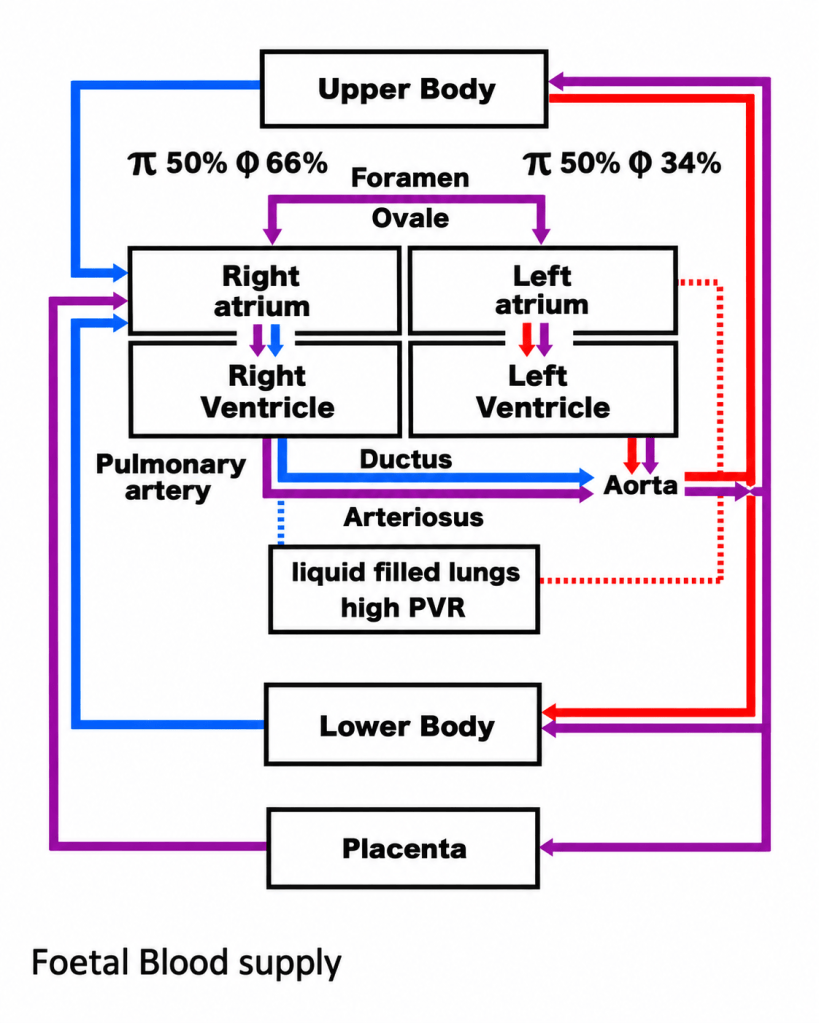
PVR is high and systemic resistance is relatively low.
After birth:
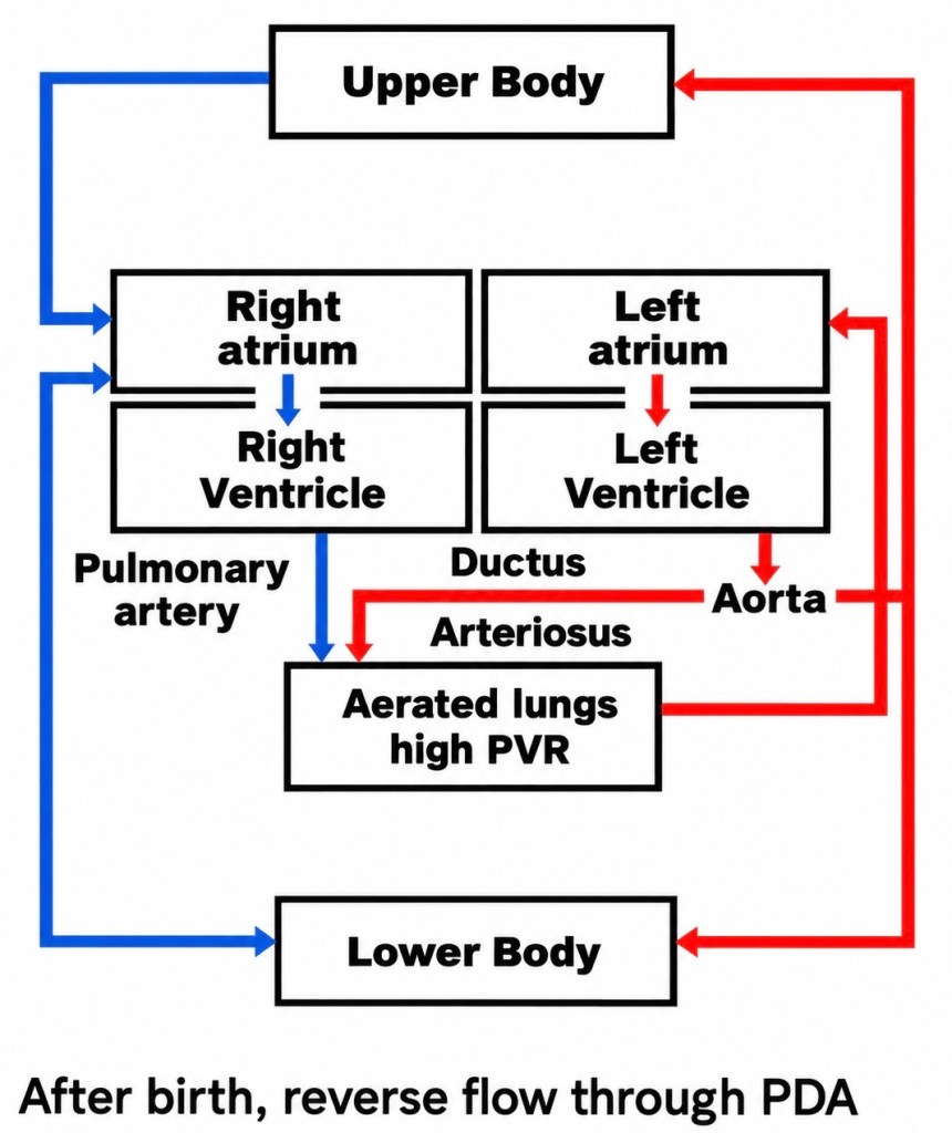
PVR falls while systemic vascular resistance rises.
Blood returning from the newly expanded lungs increases left atrial pressure.
Left atrial pressure subsequently exceeds right atrial pressure, pushing the flap of the septum primum against the septum secundum and functionally closing the foramen ovale.
Flow through the ductus arteriosus also changes direction shortly after birth as systemic arterial pressure becomes greater than pulmonary arterial pressure. Increasing oxygen tension and falling prostaglandin activity promote ductal constriction. (NCBI)
An important distinction should be made between functional closure and anatomical closure.
The ductus can become functionally closed relatively quickly, while complete anatomical obliteration occurs later.
This transition from foetal to neonatal circulation explains why some congenital cardiac defects may appear relatively well tolerated before birth yet become critically important after delivery.
Certain lesions depend upon continued flow through the ductus arteriosus to maintain either pulmonary or systemic circulation.
These are described as duct-dependent lesions.
Why “Pink Babies” and “Blue Babies”?
One of the simplest ways of introducing congenital heart disease is to divide defects into “Pink Babies” and “Blue Babies.”
These are teaching terms rather than formal diagnoses, but they provide a useful starting point for understanding the underlying physiology.
A Pink Baby has sufficient oxygenated blood reaching the systemic circulation to avoid obvious central cyanosis. The problem is usually either a left-to-right shunt, causing oxygenated blood to be recirculated through the lungs, or an obstructive lesion that makes a ventricle work harder without initially reducing systemic oxygen saturation. Standard clinical classifications similarly group ASD, VSD, PDA and AVSD as acyanotic left-to-right shunts, and Aortic Stenosis, Pulmonary Stenosis and Coarctation as acyanotic obstructive lesions.
A Blue Baby has insufficiently oxygenated blood reaching the systemic circulation. This may occur because blood cannot reach the lungs normally, because oxygenated and deoxygenated blood mix abnormally, or because deoxygenated blood bypasses the pulmonary circulation and enters the systemic circulation. Tetralogy of Fallot, Transposition of the Great Arteries, Pulmonary Atresia, Tricuspid Atresia, Truncus Arteriosus and TAPVR are recognised examples of cyanotic congenital heart disease.
The distinction is deliberately simple. It describes physiology, not necessarily the anatomical diagnosis. Some defects can be pink in one patient and blue in another, or change as pulmonary vascular resistance, obstruction, ductal patency or shunt direction changes.
Acyanotic does not mean mild. A pink baby can have severe heart failure, pulmonary hypertension, systemic obstruction or airway compression without ever appearing obviously cyanosed.
Congenital Defects — Pink and Blue Babies
| Pink Babies — Usually Acyanotic | Blue Babies — Usually Cyanotic |
| Atrial Septal Defect — ASD | Tetralogy of Fallot — TOF |
| Ventricular Septal Defect — VSD | Transposition of the Great Arteries — TGA |
| Patent Ductus Arteriosus — PDA | Pulmonary Atresia |
| Atrioventricular Septal Defect — AVSD | Pulmonary Atresia + VSD + MAPCAs |
| Coarctation of the Aorta — CoA | Tricuspid Atresia |
| Aortic Stenosis | Total Anomalous Pulmonary Venous Return — TAPVR |
| Pulmonary Stenosis — mild/moderate | Truncus Arteriosus |
| Partial Anomalous Pulmonary Venous Return — PAPVR | Hypoplastic Left Heart Syndrome — HLHS* |
| Scimitar Syndrome | Ebstein Anomaly — severe forms |
| Aortopulmonary Window | Double Outlet Right Ventricle — DORV* |
| Double Aortic Arch / Vascular Ring | Critical Pulmonary Stenosis* |
| Other functionally single-ventricle lesions |
The Grey Areas
Some lesions do not fit neatly into one column.
HLHS is anatomically a severe left-sided obstructive lesion, but the newborn circulation depends upon atrial mixing and ductal systemic blood flow, and systemic oxygen saturation is typically reduced. For the physiological approach used in this series, it is therefore included with the Blue Babies. Formal classifications may instead place it among obstructive lesions while recognising that it often produces cyanosis.
DORV can be pink or blue depending upon the position of the VSD, the relationship of the great arteries and whether pulmonary outflow obstruction is present.
Pulmonary Stenosis is generally pink when mild or moderate, but critical neonatal obstruction can severely restrict pulmonary blood flow and produce cyanosis.
Coarctation is usually pink, although a critically ill neonate with duct-dependent systemic circulation may develop differential cyanosis.
MAPCAs are not themselves an intracardiac defect. They are abnormal systemic arteries supplying the lungs, most importantly in association with Pulmonary Atresia/VSD and severe TOF, and are therefore discussed within the Blue Baby section.
So the useful starting point is:
Pink Baby = oxygenation is broadly preserved, but blood is flowing in the wrong quantity, direction or against an obstruction.
Blue Baby = too little adequately oxygenated blood is reaching the systemic circulation.
And in congenital cardiac disease, the most useful question remains:
Where is the blood going — and why?
Glossary of Terms
Aortic Arch
The curved portion of the aorta connecting the ascending and descending aorta.
It gives rise to the major arteries supplying the head, neck and upper limbs.
Aortic Stenosis
Narrowing of the pathway through which blood leaves the left ventricle and enters the aorta.
The obstruction may be:
- subvalvar;
- valvar;
- or supravalvar.
Atrial Septal Defect — ASD
An abnormal communication between the right and left atria.
In most uncomplicated ASDs, blood flows from the higher-pressure left atrium to the right atrium, producing a left-to-right shunt.
Atrioventricular Septal Defect — AVSD
A congenital abnormality involving the atrial and ventricular septa and the atrioventricular valves.
AVSD may be partial, transitional or complete and is strongly associated with Trisomy 21.
Bicuspid Aortic Valve — BAV
An aortic valve formed with two functional leaflets rather than the usual three.
It may be associated with aortic stenosis, aortic regurgitation, dilation of the ascending aorta and Coarctation of the Aorta.
Cardiac Catheterisation
A procedure in which catheters are passed through blood vessels into the heart.
It may be used to measure pressures, assess oxygen saturations, define anatomy or perform interventions such as balloon dilation or device closure.
Coarctation of the Aorta — CoA
Congenital narrowing of the aorta, most commonly near the aortic isthmus close to the ductus arteriosus.
It produces obstruction to systemic blood flow rather than primarily causing abnormal oxygenation.
Cyanosis
Bluish discoloration of the lips, tongue or skin caused by an increased quantity of deoxygenated haemoglobin in systemic arterial blood.
Most “pink baby” lesions are not cyanotic under normal circumstances.
Differential Cyanosis
A difference in oxygen saturation between the upper and lower parts of the body.
This may occur in critical neonatal Coarctation when lower-body circulation depends upon blood passing through the ductus arteriosus.
Ductus Arteriosus
A normal foetal blood vessel connecting the pulmonary artery to the aorta.
It normally closes after birth.
Echocardiography
Ultrasound imaging of the heart.
It can assess cardiac anatomy, valve function, blood-flow direction, ventricular function and pressure gradients.
Eisenmenger Syndrome
A late complication of a longstanding large left-to-right shunt in which progressive pulmonary vascular disease causes pulmonary vascular resistance to rise sufficiently for the shunt to become bidirectional or right-to-left.
The patient may then develop cyanosis.
Heart Failure
A clinical condition in which the heart cannot meet the circulatory demands of the body without abnormal filling pressures or compensatory mechanisms.
In infants with large left-to-right shunts, heart failure commonly presents with tachypnoea, sweating during feeds and poor weight gain.
Left-to-Right Shunt
Abnormal movement of blood from the systemic side of the circulation to the pulmonary side.
Because the blood has already been oxygenated, systemic oxygen saturation may remain normal despite increased pulmonary blood flow.
Left Ventricular Hypertrophy — LVH
Thickening of the muscular wall of the left ventricle.
It can develop when the left ventricle must pump against increased pressure, such as in Aortic Stenosis or severe Coarctation.
Murmur
A sound produced by turbulent blood flow through the heart or great vessels.
A murmur may result from abnormal valves, septal defects, increased flow or vascular obstruction.
Patent Ductus Arteriosus — PDA
Persistence of the ductus arteriosus after it would normally have closed.
In the usual postnatal circulation, blood flows from the higher-pressure aorta into the pulmonary artery, producing a left-to-right shunt.
Partial Anomalous Pulmonary Venous Return — PAPVR
A congenital abnormality in which one or more, but not all, pulmonary veins drain into the systemic venous circulation rather than the left atrium.
This usually creates a left-to-right shunt.
Pink Baby
An informal teaching term describing a baby with congenital heart disease whose systemic oxygenation remains sufficient to prevent obvious central cyanosis.
The term does not imply that the defect is minor.
A “pink” baby may still have severe heart failure, pulmonary hypertension or critical systemic obstruction.
Pulmonary Artery Banding
A surgical procedure in which a band is placed around the pulmonary artery to deliberately restrict excessive pulmonary blood flow.
It is occasionally used as part of staged management for complex congenital cardiac disease.
Pulmonary Hypertension
Abnormally elevated pressure within the pulmonary arterial circulation.
Longstanding excessive pulmonary blood flow can eventually lead to pulmonary vascular disease and pulmonary hypertension.
Pulmonary Over-Circulation
Excessive blood flow through the lungs.
It commonly occurs in large left-to-right shunts such as significant VSD, PDA or AVSD.
Pulmonary Stenosis
Narrowing of the pathway between the right ventricle and pulmonary arteries.
The obstruction may be:
- subvalvar;
- valvar;
- or supravalvar.
Mild and moderate disease is usually acyanotic, while critical obstruction can significantly restrict pulmonary blood flow and produce cyanosis.
Pulmonary Vascular Resistance — PVR
The resistance encountered by blood passing through the pulmonary circulation.
PVR is normally high before birth and falls rapidly after the lungs expand.
Qp:Qs Ratio
A comparison of pulmonary blood flow (Qp) with systemic blood flow (Qs).
A ratio greater than 1:1 indicates that more blood is passing through the pulmonary circulation than the systemic circulation, as occurs with significant left-to-right shunting.
Radiofemoral Delay
A delay between the palpable pulse in the radial artery at the wrist and the femoral artery in the groin.
It is a classic clinical sign associated with Coarctation of the Aorta.
Right Ventricular Hypertrophy — RVH
Thickening of the right ventricular muscle caused by increased pressure workload.
It can develop in conditions such as significant Pulmonary Stenosis or pulmonary hypertension.
Scimitar Syndrome
A form of Partial Anomalous Pulmonary Venous Return usually involving abnormal drainage of right pulmonary veins towards the inferior vena cava.
It may also be associated with right lung hypoplasia and abnormal systemic arterial supply to the right lung.
Septum
A muscular or fibrous wall separating two chambers of the heart.
The atrial septum separates the atria and the ventricular septum separates the ventricles.
Shunt
Abnormal movement of blood between two parts of the circulation.
Shunts may be:
- left-to-right;
- right-to-left;
- or bidirectional.
Systemic Vascular Resistance — SVR
The resistance encountered by blood flowing through the systemic circulation.
It represents the afterload against which the left ventricle normally pumps.
Trisomy 21
A chromosomal condition caused by an additional copy of chromosome 21.
It is particularly associated with Atrioventricular Septal Defect.
Vascular Ring
An abnormal arrangement of the aortic arch or its branches that surrounds and potentially compresses the trachea and oesophagus.
Examples include Double Aortic Arch.
The problem is principally one of airway and oesophageal compression, rather than abnormal intracardiac blood flow.
Ventricular Septal Defect — VSD
An abnormal communication between the right and left ventricles.
After birth, blood usually flows from the higher-pressure left ventricle into the right ventricle.
Large defects can cause substantial pulmonary over-circulation and heart failure.
Volume Overload
An increase in the amount of blood a cardiac chamber must handle.
For example, ASD causes right atrial and right ventricular volume overload, while a large VSD or PDA can produce left-sided volume overload because of increased pulmonary venous return.
Pressure Overload
An increase in the pressure against which a ventricle must pump.
Examples include:
- left ventricular pressure overload in Aortic Stenosis or Coarctation;
- right ventricular pressure overload in Pulmonary Stenosis.
Device Closure
Catheter-based closure of a congenital communication using an implanted occlusion device.
It may be suitable for selected ASDs, PDAs and some VSDs.
Balloon Valvuloplasty
A catheter procedure in which a balloon is inflated across a narrowed valve to increase the size of the opening.
It is commonly used for suitable congenital Pulmonary Stenosis and Aortic Stenosis.
Balloon Angioplasty
Catheter-based dilation of a narrowed blood vessel using an inflatable balloon.
It may be used in selected cases of Coarctation or recurrent Coarctation.
Stent
A metal scaffold placed within a blood vessel to hold it open.
Stents may be used in selected older children and adults with Coarctation of the Aorta.
Pulmonary-to-Systemic Flow Balance
The relationship between blood flowing through the pulmonary and systemic circulations.
Many acyanotic congenital defects produce excessive pulmonary flow while maintaining adequate systemic oxygen saturation.
Pulmonary Vascular Disease
Structural changes within the pulmonary arteries caused by persistently abnormal pressure or flow.
Severe irreversible pulmonary vascular disease can eventually prevent safe closure of a longstanding cardiac shunt.
Central Cyanosis
Bluish discoloration involving the tongue and central mucous membranes caused by reduced systemic arterial oxygen saturation.
It should be distinguished from peripheral cyanosis caused by slow circulation or cold extremities.
Acyanotic Does Not Mean Mild
An important principle in congenital cardiac disease.
A patient may remain completely “pink” while suffering severe:
- heart failure;
- pulmonary hypertension;
- systemic obstruction;
- ventricular hypertrophy;
- or progressive pulmonary vascular disease.
The pink/blue distinction describes oxygenation, not the overall severity of the heart defect.

