Thoracic surgery covers an enormous range of procedures involving the lungs, pleura, mediastinum, chest wall and associated structures.
At one end of the spectrum, a surgeon may obtain a small lymph-node biopsy through an endoscope.
At the other, an entire lung may be removed through an open thoracotomy.
Modern thoracic surgery has increasingly moved towards minimally invasive approaches such as Video-Assisted Thoracoscopic Surgery — VATS — and Robotic-Assisted Thoracic Surgery — RATS — but conventional open thoracotomy remains essential for some operations.
Understanding thoracic surgery therefore requires separating two related questions:
How does the surgeon gain access to the chest?
and
What operation is performed once access has been obtained?
A lobectomy, for example, describes what is removed.
VATS, RATS or thoracotomy describes how the surgeon gets there.
That distinction is important.
The Thoracic Cavity
The lungs occupy the right and left pleural cavities.
Between them lies the mediastinum, containing structures including:
- the heart;
- great vessels;
- trachea;
- main bronchi;
- oesophagus;
- lymphatic structures;
- vagus and phrenic nerves;
- thymic tissue;
- and mediastinal lymph nodes.
Thoracic surgery may consequently involve much more than simply removing lung tissue.
Procedures may be undertaken for:
- lung cancer;
- pulmonary nodules;
- emphysema;
- bullous lung disease;
- pneumothorax;
- pleural disease;
- mediastinal tumours;
- lymph-node biopsy;
- infection;
- bronchiectasis;
- chest-wall deformity;
- oesophageal disease;
- trauma;
- and transplantation.
The operative approach is selected according to the pathology, anatomical location, extent of surgery required and condition of the patient.
Anatomy of the Respiratory System
Before looking at thoracic surgery, double-lumen tubes or single-lung ventilation, it helps to follow the respiratory tract from the point at which the anaesthetist first sees the airway down to the microscopic level where gas exchange actually occurs.
The pathway is:
vocal cords → trachea → carina → main bronchi → lobar and segmental bronchi → bronchioles → alveoli
Each part becomes directly relevant during thoracic anaesthesia.
The Vocal Cords
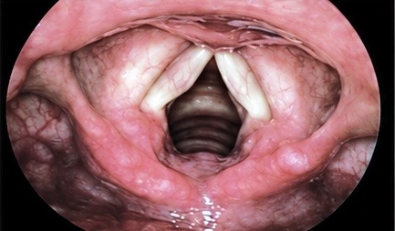
The vocal cords, or vocal folds, sit within the larynx and form the entrance to the lower respiratory tract.
When viewed during laryngoscopy, the cords usually appear as two pale structures surrounding the dark opening of the glottis.
During normal inspiration the cords abduct, allowing air to enter the trachea.
During phonation they move towards one another and vibrate as air passes between them.
For the anaesthetic practitioner, their importance is immediate.
Any tracheal tube must pass between the vocal cords before entering the trachea.
This is particularly relevant in thoracic anaesthesia because double-lumen endotracheal tubes are larger, more rigid and more curved than conventional single-lumen tubes.
Careful passage through the cords is therefore important to avoid:
- vocal-cord trauma;
- mucosal injury;
- oedema;
- arytenoid injury;
- postoperative sore throat;
- and, rarely, vocal-cord dysfunction.
Once the tube has passed through the vocal cords, the next major anatomical landmark is the distal trachea.
The Trachea and Carina
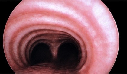
The trachea is supported by a series of C-shaped cartilaginous rings which maintain airway patency.
It extends from the larynx through the neck and upper mediastinum before dividing into the right and left main bronchi.
This division is marked by the:
Carina
The carina appears bronchoscopically as a central ridge separating the entrances to the right and left main bronchi.
It is one of the most important landmarks in thoracic anaesthesia.
When positioning a double-lumen tube, the carina provides the reference point from which the anaesthetic team determines whether the bronchial component of the tube is correctly positioned.
With a conventional left-sided double-lumen tube, the bronchial lumen passes into the left main bronchus, with the bronchial cuff sitting just below the carina.
If the tube is too shallow, the bronchial cuff may sit partly within the trachea.
If it is advanced too far, it may obstruct the left upper- or lower-lobe bronchi.
For this reason, correct position is usually confirmed with fibreoptic bronchoscopy.
In camera-equipped double-lumen tubes, the relationship between the tube and the carina may also be viewed continuously.
The carina is therefore not simply an anatomical division.
During thoracic anaesthesia it becomes a crucial positioning landmark.
The Bronchial Tree
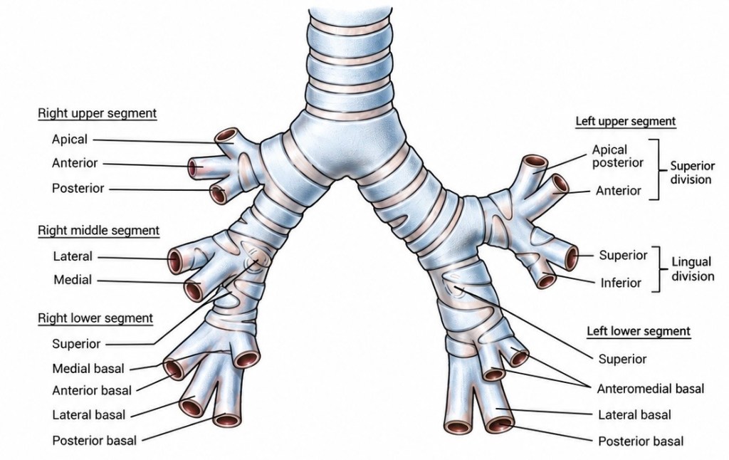
Below the carina, the airways divide repeatedly to distribute inspired gas throughout the lungs.
The first major division is into the:
right main bronchus
and
left main bronchus.
These are anatomically different.
The right main bronchus is generally:
- shorter;
- wider;
- and more vertical.
The left main bronchus is:
- longer;
- narrower;
- and more oblique.
This is one of the reasons left-sided double-lumen tubes are usually preferred for routine lung isolation.
The longer left main bronchus provides a more forgiving position for the bronchial cuff.
The Right Lung
The right lung contains three lobes:
- upper;
- middle;
- lower.
Each lobe is divided further into bronchopulmonary segments.
Right Upper Lobe
The principal segments are:
- apical;
- anterior;
- posterior.
Right Middle Lobe
The principal segments are:
- lateral;
- medial.
Right Lower Lobe
The principal segments are:
- superior;
- medial basal;
- anterior basal;
- lateral basal;
- posterior basal.
The right upper-lobe bronchus arises relatively close to the carina.
This is one of the major challenges when using a right-sided double-lumen tube.
The tube must isolate the right main bronchus while simultaneously allowing ventilation through an opening aligned with the right upper-lobe bronchus.
Even relatively small tube movement can compromise that alignment.
The Left Lung
The left lung has two lobes:
- upper;
- lower.
The left upper lobe also contains the lingula, which is anatomically comparable in some respects with the right middle lobe.
Left Upper Lobe
The principal segments include:
Superior division
- apicoposterior;
- anterior.
Lingular division
- superior lingular;
- inferior lingular.
Left Lower Lobe
The principal segments include:
- superior;
- anteromedial basal;
- lateral basal;
- posterior basal.
There is some normal anatomical variation in segmental arrangement.
This becomes particularly relevant during anatomical segmentectomy.
Bronchopulmonary Segments
A bronchopulmonary segment is a relatively discrete anatomical unit of lung supplied by its own:
- segmental bronchus;
- pulmonary arterial branch;
- and associated vascular anatomy.
These segmental boundaries have major surgical importance.
They allow surgeons to remove an individual segment while preserving the remainder of the lobe.
This is the basis of anatomical segmentectomy.
It also explains why understanding bronchial anatomy is essential during:
- thoracic surgery;
- bronchoscopy;
- lung isolation;
- bronchial blocker placement;
- and double-lumen tube positioning.
The bronchial tree is therefore not simply a set of branching airways.
It is also the anatomical map used during thoracic surgery.
From Bronchi to Bronchioles
As the bronchial tree divides repeatedly, the airways become progressively smaller.
Larger bronchi are supported by cartilage.
Further distally, the airways become bronchioles, which lack the same cartilaginous support.
The pathway progresses through:
bronchi → bronchioles → terminal bronchioles → respiratory bronchioles → alveolar ducts → alveolar sacs → alveoli
At this point the respiratory system moves from conducting air to exchanging gases.
The Alveoli
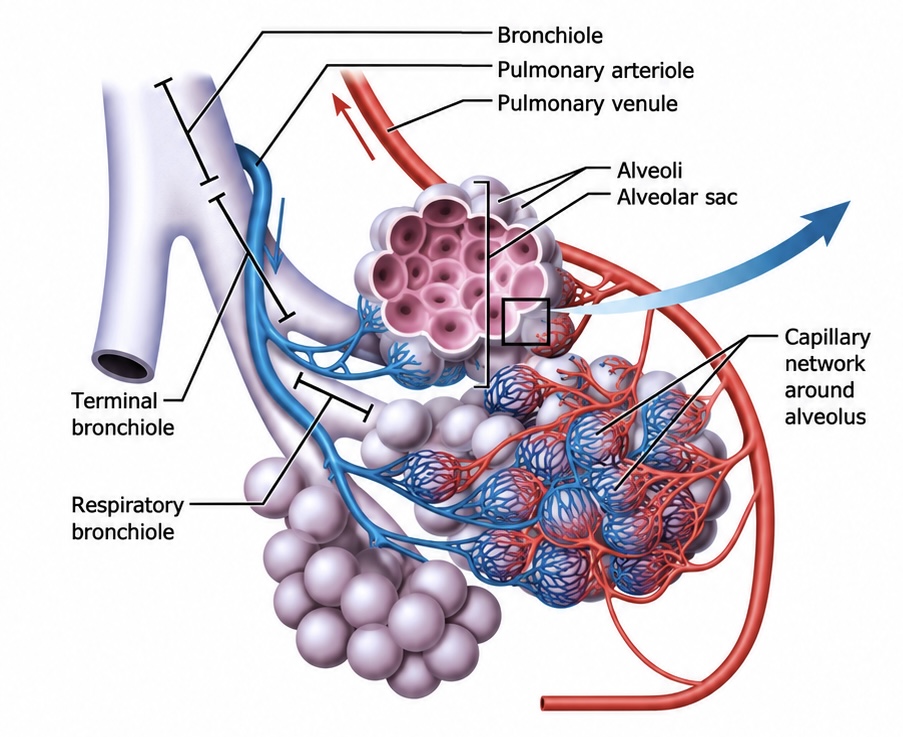
The alveoli are the microscopic gas-exchange units of the lungs.
There are several hundred million alveoli in the adult respiratory system, creating an enormous surface area across which oxygen and carbon dioxide can move.
Each alveolus is surrounded by a dense capillary network.
The barrier between alveolar gas and pulmonary capillary blood is extremely thin.
This allows oxygen to travel:
alveolus → pulmonary capillary blood
while carbon dioxide travels in the opposite direction:
blood → alveolus
The efficiency of this process depends upon both ventilation and perfusion.
Type I Pneumocytes
Most of the gas-exchange surface of the alveolus is formed by Type I pneumocytes.
These cells are extremely thin.
Their structure minimises the distance through which oxygen and carbon dioxide must diffuse.
Type II Pneumocytes and Surfactant
Type II pneumocytes have several functions, but one of the most important is the production of pulmonary surfactant.
Surfactant is a mixture of phospholipids and proteins that coats the inner surface of the alveolus.
It reduces surface tension and helps prevent alveolar collapse during expiration.
Without adequate surfactant, the alveoli would require considerably more pressure to remain open.
Surfactant therefore helps:
- maintain alveolar stability;
- reduce the work of breathing;
- improve lung compliance;
- and reduce end-expiratory alveolar collapse.
Alveolar Macrophages
The alveoli also contain alveolar macrophages.
These cells form part of the lung’s immune and housekeeping system.
They remove:
- inhaled particles;
- pathogens;
- cellular debris;
- and excess surfactant.
Their function is influenced by Granulocyte-Macrophage Colony-Stimulating Factor — GM-CSF.
This becomes particularly relevant in Pulmonary Alveolar Proteinosis.
In autoimmune PAP, antibodies interfere with GM-CSF signalling.
The alveolar macrophages become less effective at clearing surfactant.
Surfactant-rich material then accumulates within the alveoli.
The result is impaired gas exchange.
The anatomy shown in this image therefore becomes directly relevant to understanding why PAP occurs and why whole-lung lavage can be effective.
Ventilation and Perfusion
Gas exchange requires both:
Ventilation
Air reaching the alveolus.
and
Perfusion
Blood reaching the surrounding capillary network.
When an alveolus is ventilated and perfused appropriately, gas exchange can occur efficiently.
If blood reaches an area of lung that is not being ventilated, that blood passes through the pulmonary circulation without being fully oxygenated.
This contributes to a:
Right-to-left intrapulmonary shunt.
This is exactly what occurs when one lung is deliberately collapsed during single-lung ventilation.
Blood continues to flow through the non-ventilated lung.
The body responds through hypoxic pulmonary vasoconstriction, reducing blood flow to the poorly ventilated lung and diverting more blood towards the lung that remains ventilated.
This means the microscopic anatomy of the alveolus becomes directly relevant to what happens during major thoracic surgery.
From the Vocal Cords to the Alveoli
These four images effectively demonstrate the entire route followed by inspired gas.
The anaesthetist first sees the:
vocal cords.
The trachea then leads to the:
carina.
The carina divides into the:
right and left bronchial trees.
These progressively divide into increasingly smaller airways until inspired gas finally reaches the:
alveoli.
That anatomical journey explains much of thoracic anaesthesia.
The vocal cords determine access to the airway.
The carina determines double-lumen tube position.
The bronchial tree determines which lung, lobe or segment can be isolated.
And the alveoli determine whether the patient can continue to exchange oxygen and carbon dioxide when one lung is deliberately removed from ventilation.
Understanding those structures is therefore the foundation for understanding:
double-lumen tubes, bronchial blockers, single-lung ventilation and thoracic surgery itself.
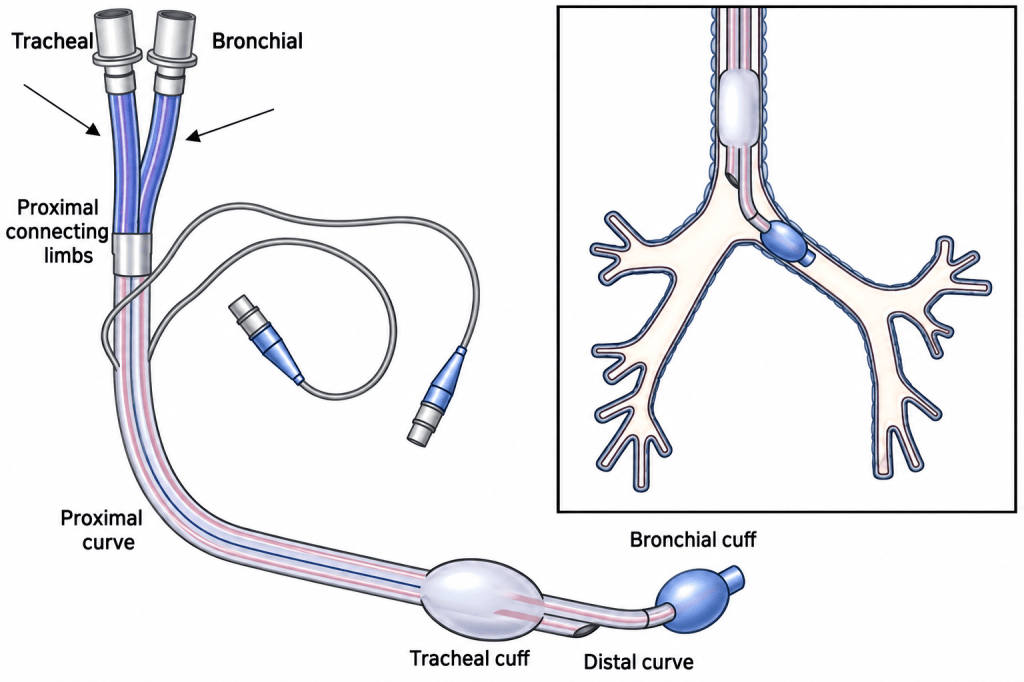
Lung Isolation and Single-Lung Ventilation
One of the defining features of thoracic anaesthesia is lung isolation.
For many intrathoracic procedures the surgeon needs the operative lung to remain still and collapsed.
Trying to operate on a fully inflated lung that expands and contracts with every breath would make precise dissection extremely difficult.
The anaesthetic team therefore mechanically separates the two lungs.
This is most commonly achieved using a:
double-lumen endobronchial tube — DLT
or, in selected circumstances, a:
bronchial blocker.
The operative lung is allowed to collapse while the opposite lung is mechanically ventilated.
This is known as:
Single-Lung Ventilation — SLV.
Single-lung ventilation provides surgical exposure but creates significant physiological consequences.
Blood continues flowing through the non-ventilated lung, creating a right-to-left intrapulmonary shunt.
Hypoxic pulmonary vasoconstriction helps compensate by diverting pulmonary blood flow away from the non-ventilated lung towards the ventilated lung.
The anaesthetic team must balance adequate oxygenation and carbon-dioxide elimination against protection of the single ventilated lung.
Single-lung ventilation therefore links the surgical and anaesthetic components of thoracic surgery.
Surgical Access to the Chest
There are several major approaches.
These can broadly be divided into:
- open surgery;
- minimally invasive thoracoscopic surgery;
- robotic-assisted surgery;
- and specialised approaches to the mediastinum.
Thoracotomy
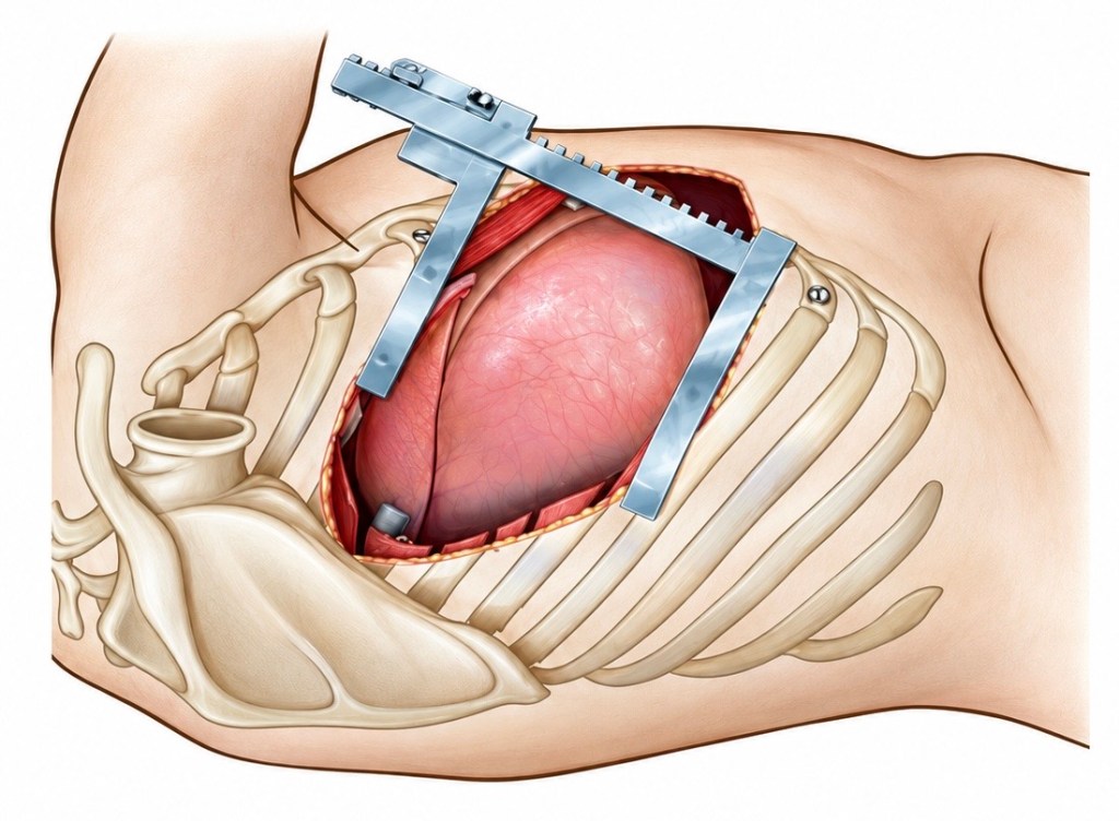
A thoracotomy is an open surgical incision into the chest.
For many decades this was the standard approach for major pulmonary surgery.
A traditional posterolateral thoracotomy involves a substantial incision extending around the lateral chest wall towards the back.
Depending upon the precise approach, chest-wall muscles may be divided or mobilised, including:
- latissimus dorsi;
- serratus anterior;
- and portions of the pectoral musculature.
The intercostal space is opened and the ribs separated using a rib spreader.
This provides the surgeon with direct access to the thoracic cavity.
Advantages of Thoracotomy
The great advantage is exposure.
The surgeon can directly see and manipulate the lung, hilum, pulmonary vessels, mediastinum and chest wall.
Thoracotomy remains particularly important when dealing with:
- very large tumours;
- locally invasive malignancy;
- major vascular involvement;
- complex airway reconstruction;
- major bleeding;
- chest-wall resection;
- difficult adhesions;
- and situations where minimally invasive surgery has to be converted to an open operation.
The Price of Exposure
Thoracotomy is highly effective but physiologically expensive.
The incision, intercostal trauma and rib spreading can produce considerable postoperative pain.
Pain inhibits deep inspiration and coughing.
This can contribute to:
- reduced tidal volume;
- atelectasis;
- retained secretions;
- sputum retention;
- pneumonia;
- impaired mobilisation;
- and postoperative respiratory failure.
Effective analgesia is therefore an integral part of thoracic anaesthesia rather than merely postoperative comfort.
Techniques can include regional blocks, paravertebral techniques, fascial-plane blocks, epidural analgesia and multimodal systemic analgesia.
Video-Assisted Thoracoscopic Surgery — VATS
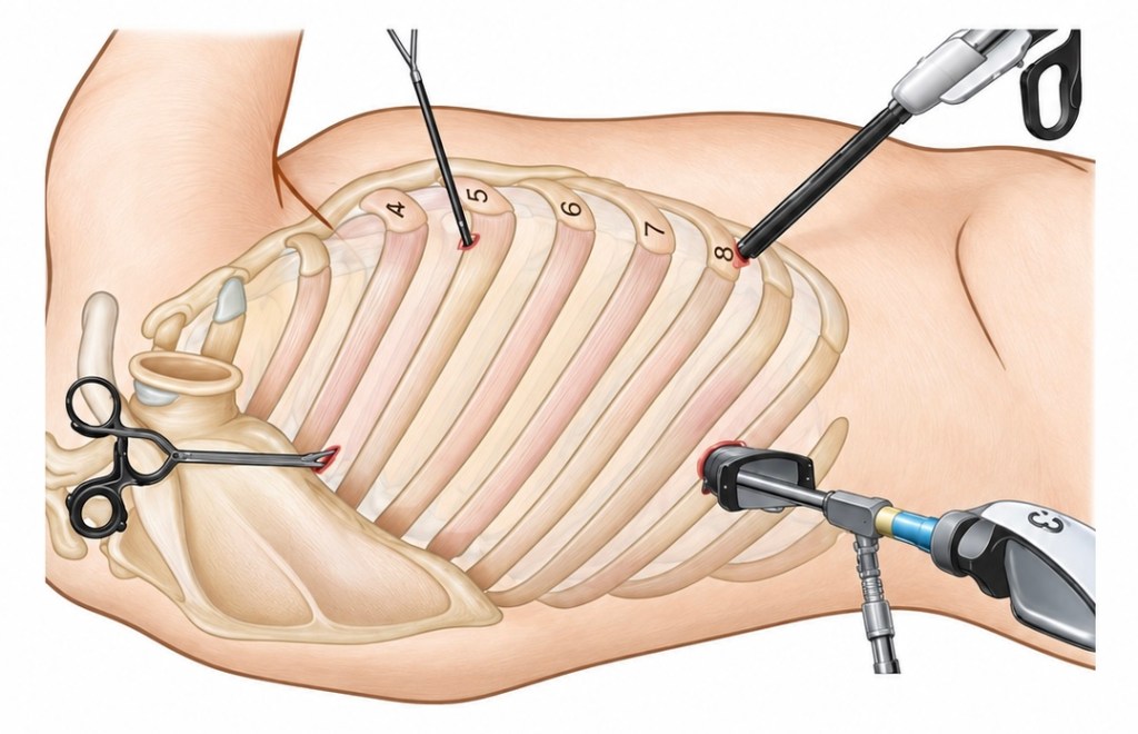
The development of Video-Assisted Thoracoscopic Surgery transformed thoracic surgery.
Rather than making one large incision and spreading the ribs widely, a camera and surgical instruments are inserted through much smaller chest-wall incisions.
The surgeon views the operation on a high-definition monitor.
Traditionally VATS involved three or four ports.
A camera port, commonly around 5–10 mm, may be positioned in the lower lateral chest.
A somewhat larger utility incision provides access for instruments and retrieval of tissue.
Other ports allow introduction of graspers, dissectors, staplers and suction.
The exact configuration depends upon:
- the operation;
- the lobe involved;
- surgeon preference;
- patient anatomy;
- and whether a multiportal or uniportal technique is being used.
Uniportal VATS
Modern VATS increasingly includes uniportal surgery.
Rather than using several separate access points, the camera and surgical instruments are passed through a single intercostal incision.
The basic surgical objective remains the same, but the approach attempts to minimise chest-wall trauma further.
What Can Be Done Through VATS?
VATS is no longer restricted to minor diagnostic procedures.
Modern thoracic surgeons can perform major anatomical resections through thoracoscopic approaches, including:
- wedge resection;
- segmentectomy;
- lobectomy;
- bullectomy;
- pleural procedures;
- mediastinal procedures;
- sympathectomy;
- decortication;
- and selected more complex pulmonary resections.
NICE recommends lobectomy for appropriate patients with operable non-small-cell lung cancer and explicitly recognises either an open or thoracoscopic approach. (Nice)
Advantages of VATS
Compared with conventional open thoracotomy, minimally invasive thoracic surgery avoids extensive rib spreading and generally produces less chest-wall trauma.
Potential advantages include:
- reduced postoperative pain;
- earlier mobilisation;
- shorter hospital stay;
- reduced impairment of respiratory mechanics;
- smaller surgical wounds;
- and faster functional recovery.
But a smaller incision should not create the impression that the operation itself is necessarily minor.
A VATS lobectomy remains a major anatomical lung resection.
The pulmonary artery, pulmonary vein and lobar bronchus still have to be dissected and divided.
The risk of major haemorrhage remains.
A minimally invasive approach changes access to the operation.
It does not abolish the potential seriousness of the operation itself.
Conversion from VATS to Thoracotomy
Every practitioner involved in major VATS surgery should understand one essential point:
A VATS operation can become an open thoracotomy very quickly.
Reasons include:
- major pulmonary arterial bleeding;
- unexpected anatomy;
- difficult hilar dissection;
- dense adhesions;
- inability to obtain adequate surgical margins;
- equipment failure;
- or inability to progress safely thoracoscopically.
Conversion should not be regarded simply as a failed VATS operation.
It is a safety strategy.
For the theatre and anaesthetic team, however, it means preparation must reflect the potential operation, not merely the size of the initial skin incision.
Robotic-Assisted Thoracic Surgery — RATS
The next major development has been Robotic-Assisted Thoracic Surgery — RATS.
The word robotic can be slightly misleading.
The robot does not independently perform the operation.
The surgeon controls the instruments from a console.
Robotic systems provide:
- high-definition three-dimensional vision;
- articulated instruments;
- tremor filtration;
- considerable instrument dexterity;
- and fine manipulation within confined anatomical spaces.
The Society of Thoracic Surgeons describes robotic anatomical lung resection as a minimally invasive approach offering enhanced 3-D visualisation and increased instrument dexterity. (consult.sts.org)
How RATS Works
Several ports are placed through the chest wall.
Robotic instrument arms are then docked to the ports.
The surgeon operates from a console rather than standing immediately adjacent to the patient.
A bedside assistant remains scrubbed at the operating table and may:
- change instruments;
- introduce stapling devices;
- apply suction;
- retrieve specimens;
- assist with vascular control;
- and respond immediately during an emergency.
This last point matters considerably.
If catastrophic haemorrhage occurs, the robotic system has to be undocked sufficiently to permit emergency open access.
The entire team therefore needs a clearly understood emergency conversion strategy.
VATS or RATS?
The two techniques should not necessarily be viewed as rivals.
Both are forms of minimally invasive thoracic surgery.
VATS gives the surgeon a camera-based thoracoscopic view and uses long instruments introduced directly through the ports.
RATS introduces articulated robotic instruments under console control.
Choice depends upon:
- local expertise;
- surgeon training;
- equipment availability;
- patient anatomy;
- disease;
- and the procedure being performed.
Both may ultimately achieve the same anatomical operation.
Mediastinoscopy
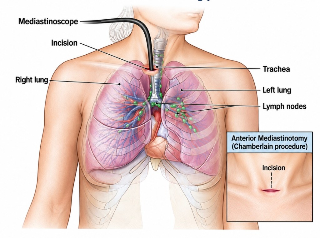
Thoracic surgery is not limited to entering the pleural cavity.
Mediastinoscopy provides access to the mediastinum.
Traditionally, cervical mediastinoscopy has played an important role in investigating and staging lung cancer.
A small incision is made just above the suprasternal notch.
Dissection proceeds into the pretracheal plane.
A mediastinoscope is advanced along this surgically created pathway towards the carina.
This gives access to mediastinal lymph nodes, particularly stations:
2, 4 and 7.
Tissue can then be obtained for histological examination.
Mediastinoscopy in Modern Practice
The role of mediastinoscopy has changed considerably since the original 2019 material was written.
Endobronchial ultrasound-guided transbronchial needle aspiration — EBUS-TBNA — and endoscopic ultrasound — EUS — now allow many mediastinal lymph nodes to be sampled without surgical mediastinoscopy.
NICE recommends EBUS-TBNA and EUS-FNA as key invasive staging techniques and notes that they can provide diagnosis and staging more rapidly and with greater patient acceptability than surgical investigation. (Nice)
Mediastinoscopy nevertheless remains important.
NICE recommends considering surgical mediastinal staging when EBUS or EUS is negative but clinical suspicion of mediastinal malignancy remains high and the result would alter treatment. (Nice)
So mediastinoscopy has not disappeared.
Its role has become more selective.
Anterior Mediastinotomy — The Chamberlain Procedure
Another surgical route to the mediastinum is the anterior mediastinotomy, often called the Chamberlain procedure.
A small anterior chest-wall incision provides access to structures that may be less accessible by conventional cervical mediastinoscopy, particularly the anterior mediastinum and aortopulmonary window.
Again, modern endoscopic and image-guided staging has reduced the frequency with which these surgical diagnostic approaches are required.
Once Inside the Chest — What Can Be Removed?
The surgical approach describes how the thoracic cavity is accessed.
The pulmonary resection describes what lung tissue is actually removed.
There is a progression from very limited removal of tissue through to removal of an entire lung.
Bullectomy
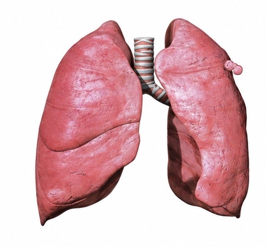
A bulla is an enlarged air-filled space within the lung resulting from destruction of normal pulmonary tissue.
Bullae are particularly associated with emphysema.
When very large, a bulla can occupy a substantial proportion of the hemithorax and compress relatively healthier surrounding lung.
A bullectomy removes the bulla.
The objective is not simply removal of an abnormal airspace.
Removing a large non-functioning bulla can allow previously compressed lung to re-expand and improve respiratory mechanics.
Bullous disease creates particular anaesthetic concerns because positive-pressure ventilation may expand a large bulla and potentially contribute to rupture or pneumothorax.
Wedge Resection
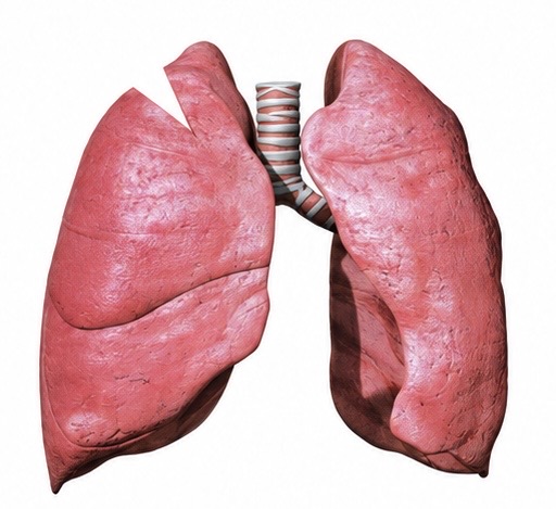
A wedge resection removes a relatively small wedge-shaped portion of lung.
It is a non-anatomical resection.
That distinction is important.
The surgeon removes the lesion together with an appropriate margin of surrounding lung but does not dissect and divide the individual segmental bronchovascular structures.
Wedge resection may be used for:
- diagnostic lung biopsy;
- pulmonary nodules;
- selected peripheral tumours;
- metastatic deposits;
- and selected patients where preservation of lung tissue is especially important.
Segmentectomy
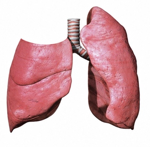
A segmentectomy is fundamentally different.
Each lung lobe consists of anatomically identifiable bronchopulmonary segments with their own segmental bronchial and vascular supply.
During anatomical segmentectomy the surgeon identifies and divides the structures supplying a specific segment and removes that segment while preserving the remainder of the lobe.
It therefore sits between wedge resection and lobectomy.
Segmentectomy has become increasingly important in modern lung cancer surgery, particularly for selected small peripheral cancers where oncological clearance can be achieved while preserving more functional lung tissue.
Lobectomy
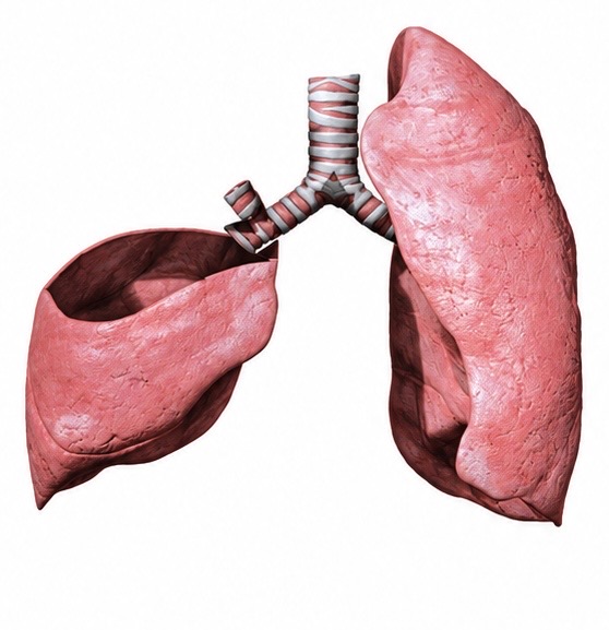
A lobectomy removes one complete lobe.
The right lung contains:
- upper lobe;
- middle lobe;
- lower lobe.
The left contains:
- upper lobe;
- lower lobe.
The operation requires anatomical division of the structures supplying that lobe:
- pulmonary artery branches;
- pulmonary veins;
- and lobar bronchus.
The lobe is then removed.
Lobectomy remains an important standard surgical treatment for suitable patients with resectable non-small-cell lung cancer. NICE recommends lobectomy in patients fit for surgery where curative treatment is appropriate. (Nice)
It may be performed through:
thoracotomy, VATS or RATS.
Again, the operation and the approach are separate concepts.
Bilobectomy
Because the right lung has three lobes, two adjacent lobes can occasionally need removal.
This is termed a bilobectomy.
Examples include:
- right upper and middle lobectomy;
- or right middle and lower lobectomy.
The procedure removes more functioning lung than a standard lobectomy but preserves the remaining right-sided lobe.
Sleeve Resection
Sometimes a tumour involves the origin of a lobar bronchus.
Historically this might have required pneumonectomy.
A sleeve resection can sometimes avoid that.
The involved lobe and a short segment of bronchus are removed.
The remaining distal airway is then reconnected to the proximal bronchus.
The principle is similar to cutting a damaged section from a pipe and reconnecting the two healthy ends.
More complex sleeve resections may also involve pulmonary arterial reconstruction.
The objective is to achieve adequate oncological clearance while preserving functioning lung whenever possible.
NICE recommends more extensive procedures such as bronchoangioplastic surgery, bilobectomy or pneumonectomy when required to achieve clear surgical margins. (Nice)
Pneumonectomy
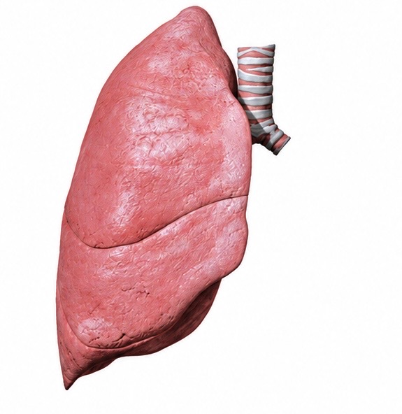
A pneumonectomy removes an entire lung.
It is one of the most physiologically significant operations in thoracic surgery.
The main pulmonary artery and veins supplying the lung are divided.
The main bronchus is divided and closed.
The entire lung is removed.
From that point onwards, all pulmonary gas exchange must ultimately be provided by the remaining lung.
Pneumonectomy is therefore reserved for situations in which adequate treatment cannot be achieved with a lesser resection.
Modern thoracic surgery generally seeks to preserve functioning lung whenever oncologically and technically possible.
What Happens to the Empty Hemithorax?
One might imagine that removing an entire lung leaves a permanent empty space filled with air.
It does not.
Following pneumonectomy, the operated hemithorax gradually fills with fluid.
Over time:
- the diaphragm rises;
- the mediastinum shifts towards the operated side;
- the remaining lung undergoes compensatory expansion;
- and the thoracic anatomy progressively remodels.
Fluid management following pneumonectomy is particularly important because the patient has only one remaining lung and excessive fluid administration may contribute to serious postoperative pulmonary complications.
Lung Volume Reduction Surgery
Not all pulmonary resection is performed for cancer.
Lung Volume Reduction Surgery — LVRS — removes severely emphysematous, poorly functioning areas of lung.
This sounds counterintuitive.
Why remove lung tissue from a patient who already has severe lung disease?
Because severely hyperinflated emphysematous lung can mechanically disadvantage the remaining healthier lung and flatten the diaphragm.
Removing carefully selected diseased areas can allow the remaining lung and diaphragm to function more effectively.
The objective is therefore not simply to create a smaller lung.
It is to create a mechanically more efficient respiratory system.
Endobronchial Valves — Surgery Without Resection
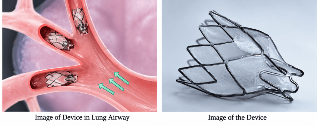
Some patients with emphysema can undergo bronchoscopic lung-volume reduction instead.
Small one-way endobronchial valves are placed within selected bronchi.
Air and secretions can leave the targeted lobe but inspired gas cannot readily enter.
The treated lobe gradually loses volume, producing an effect analogous to surgical volume reduction without physically removing the tissue.
Patient selection is critical.
The Chartis System
The Chartis system may be used during assessment for endobronchial valve treatment.
An important correction to earlier descriptions is that Chartis does not primarily measure overall lung volume.
It assesses collateral ventilation.
Normally, lobes are separated by fissures.
However, air can sometimes travel between adjacent lobes through collateral pathways.
If significant collateral ventilation is present, blocking the normal bronchial entrance to a lobe with an endobronchial valve may fail to collapse it because air simply enters from the neighbouring lobe.
Chartis therefore helps determine whether effective lobar isolation and volume reduction are likely to occur.
Chest-Wall Surgery
Thoracic surgeons also treat chest-wall deformities.
Two important operations are the Ravitch and Nuss procedures.
Pectus Excavatum and Pectus Carinatum
Pectus excavatum produces a posterior depression of the sternum, creating the characteristic funnel-chest appearance.
Pectus carinatum produces anterior displacement of the sternum and adjacent costal cartilage.
The deformity can become more pronounced during adolescent skeletal growth.
Severe pectus deformity may have functional as well as cosmetic consequences.
The Ravitch Procedure
The Ravitch procedure is an open reconstructive operation.
Abnormal costal cartilage is exposed and resected or remodelled, allowing repositioning of the sternum.
Various modifications of the original operation now exist.
The Nuss Procedure
The Nuss procedure uses a different principle.
Rather than extensively removing abnormal cartilage, one or more curved metal bars are passed behind the sternum.
The bar is rotated into position, pushing the depressed sternum anteriorly and acting as an internal brace while the chest wall remodels.
The bar remains in place for a period before being removed at a later operation.
The Nuss technique represents one of the important developments in minimally invasive chest-wall surgery.
The Common Thread: Lung Isolation
Despite the enormous differences between a diagnostic wedge biopsy, VATS lobectomy and pneumonectomy, many major intrathoracic operations share one requirement:
the surgeon needs space.
Within the closed thoracic cavity there is very little spare room.
The lung normally expands almost completely against the chest wall.
Once the pleural cavity is entered, allowing the operative lung to collapse creates the working space required for surgery.
This explains why double-lumen tubes, bronchial blockers and single-lung ventilation are so central to thoracic anaesthesia.
The surgical approach and the anaesthetic technique cannot really be understood independently of each other.
Minimally Invasive Does Not Mean Minor Surgery
This is perhaps one of the most important concepts for anyone beginning to understand modern thoracic surgery.
A patient may arrive in theatre for a procedure involving four small puncture-like incisions.
But internally the surgeon may be:
- dissecting a pulmonary artery;
- dividing pulmonary veins;
- dividing a major bronchus;
- removing an entire lobe;
- and performing mediastinal lymph-node dissection.
The external wound may be small.
The internal operation is not.
The same principle applies to robotic surgery.
Advanced technology changes the route by which surgeons reach the pathology.
It does not change the importance of the anatomy beneath it.
The Modern Thoracic Operating Theatre
Modern thoracic surgery consequently brings together several disciplines.
The surgeon needs access to the pathology.
The anaesthetist must maintain oxygenation while deliberately allowing one lung to collapse.
The anaesthetic practitioner must understand:
- double-lumen tubes;
- bronchial anatomy;
- fibreoptic bronchoscopy;
- lung isolation;
- invasive arterial monitoring;
- major haemorrhage;
- thoracic positioning;
- single-lung ventilation;
- and the possibility of emergency conversion to open surgery.
The scrub team must be prepared for both the planned minimally invasive procedure and the possibility of major open surgery.
And everyone needs to understand what operation is actually taking place beneath the skin.
That is perhaps the simplest way of understanding adult thoracic surgery:
The approach tells us how we enter the chest.
The resection tells us what is removed.
And single-lung ventilation creates the space in which much of modern thoracic surgery becomes possible.

