Lung Isolation, Bronchoscopic Placement and the Emergency Blind Technique
Introduction
One-lung ventilation is fundamental to modern thoracic anaesthesia. By separating the two lungs, the anaesthetic team can ventilate one lung while allowing the other to collapse, giving the surgeon access to structures that would otherwise remain obscured by an inflated lung.
The double-lumen endobronchial tube, or DLT, remains one of the most effective methods of achieving lung isolation.
For nurses and Operating Department Practitioners working in thoracic surgery, however, understanding a DLT involves considerably more than knowing that the tube has two lumens.
The practitioner should understand:
- why left- and right-sided tubes are different;
- how tube size is selected;
- why a left-sided tube is generally preferred;
- how the proposed operation influences which bronchus should be intubated;
- how a DLT can be positioned using a flexible bronchoscope;
- how conventional or “blind” bronchial placement works;
- how Look, Listen and Feel can establish functional lung separation;
- why clinical assessment cannot necessarily identify anatomical malposition;
- why ventilation of the right upper lobe is critical when positioning a right-sided tube;
- how bronchial cuff pressure should be managed;
- why positioning must be reassessed after turning the patient lateral;
- what to do when one-lung ventilation suddenly deteriorates;
- and why the ideal technique in elective thoracic surgery may be very different from the approach required during major trauma.
Flexible bronchoscopy is widely regarded as the gold standard for confirming DLT position.
That does not mean every DLT must necessarily be inserted over a bronchoscope.
Modern thoracic practice combines clinical assessment and bronchoscopic assessment, because they provide different information.
Clinical assessment tells us whether the tube appears to be functioning.
Bronchoscopy tells us where the tube actually is.
Why Do We Need Lung Isolation?
During conventional ventilation both lungs expand simultaneously.
In thoracic surgery this creates an obvious problem. An inflated lung occupies a substantial proportion of the hemithorax and may obscure the structures upon which the surgeon needs to operate.
Lung isolation permits one lung to collapse while ventilation continues through the opposite lung.
This can provide:
- improved surgical exposure;
- access during thoracotomy, VATS and robotic surgery;
- controlled one-lung ventilation;
- separation of contaminated and uncontaminated lungs;
- protection of one lung from blood, pus or other material arising from the opposite lung;
- selective suction;
- and rapid alternation between one- and two-lung ventilation.
DLTs are therefore commonly used during:
- wedge resections;
- segmentectomy;
- lobectomy;
- pneumonectomy;
- oesophageal surgery;
- thoracic vascular surgery;
- and selected emergency thoracic procedures.
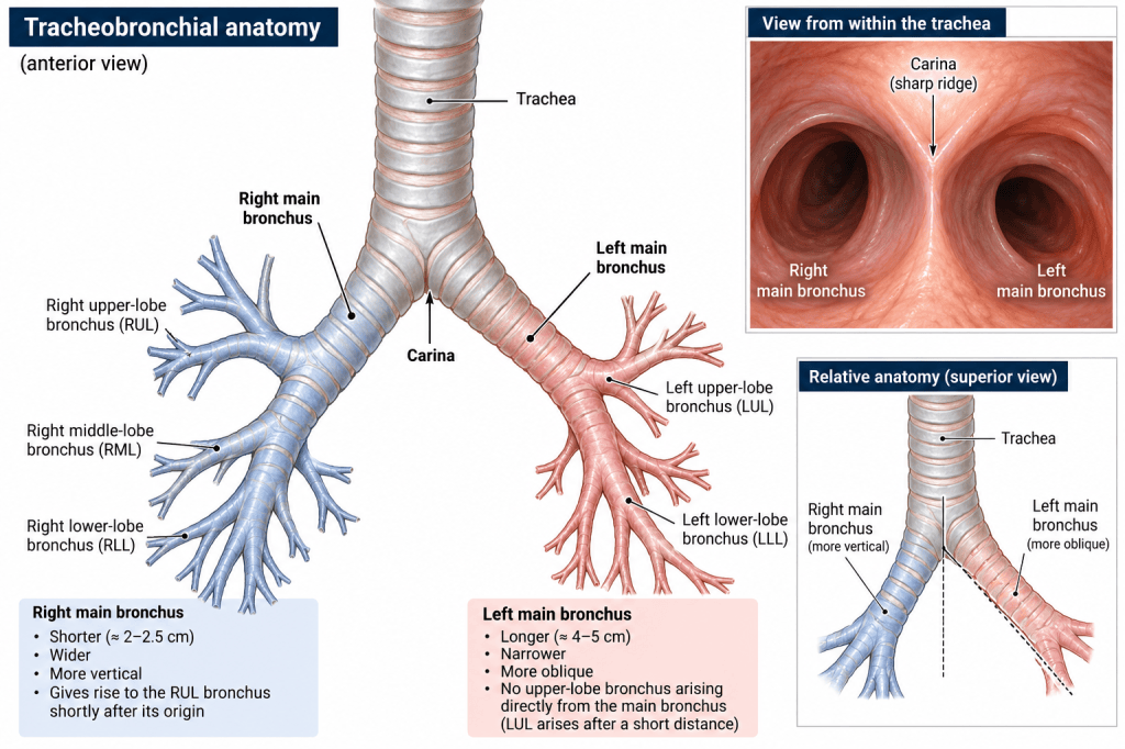
Tracheobronchial anatomy showing the carina, right main bronchus, left main bronchus and the early origin of the right upper-lobe bronchus.
What Is a Double-Lumen Tube?
A DLT effectively incorporates two airway tubes into a single device.
It contains:
A tracheal lumen, terminating within the trachea.
A bronchial lumen, extending into either the left or right main bronchus.
There are usually two cuffs:
The tracheal cuff, producing a seal within the trachea.
The bronchial cuff, producing a seal within the selected main bronchus.
The two lumens can be ventilated simultaneously or independently using the connector and clamps.
The anaesthetic team can therefore ventilate:
- both lungs;
- the right lung alone;
- or the left lung alone.
Development of the Double-Lumen Tube
Early lung-isolation devices included the Carlens tube, a left-sided tube incorporating a carinal hook.
The White tube provided a corresponding right-sided design.
The later Robertshaw tube removed the carinal hook and helped establish the configuration from which many modern disposable DLTs evolved.
Modern devices include:
- conventional PVC left-sided DLTs;
- conventional right-sided DLTs;
- reinforced or more flexible designs;
- and video DLTs containing an integrated camera.
These newer devices provide additional ways of monitoring tube position, but they do not eliminate the importance of understanding the underlying airway anatomy.
Left-Sided and Right-Sided DLTs
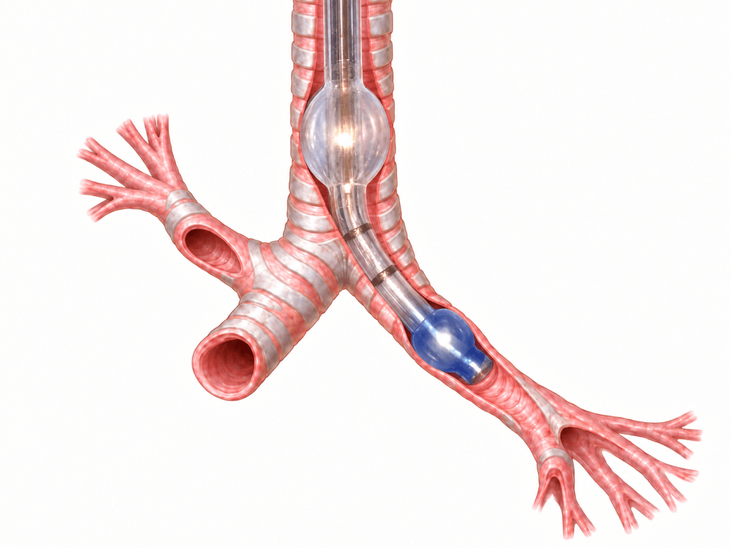
Left-Sided DLT
The bronchial lumen enters the left main bronchus.
The left main bronchus provides a relatively long section in which the bronchial cuff and tip can sit without obstructing a lobar bronchus.
This creates a useful margin of safety.
For this reason, the left-sided DLT is generally the preferred tube for adult lung isolation unless there is a specific reason why the left main bronchus should not be occupied.
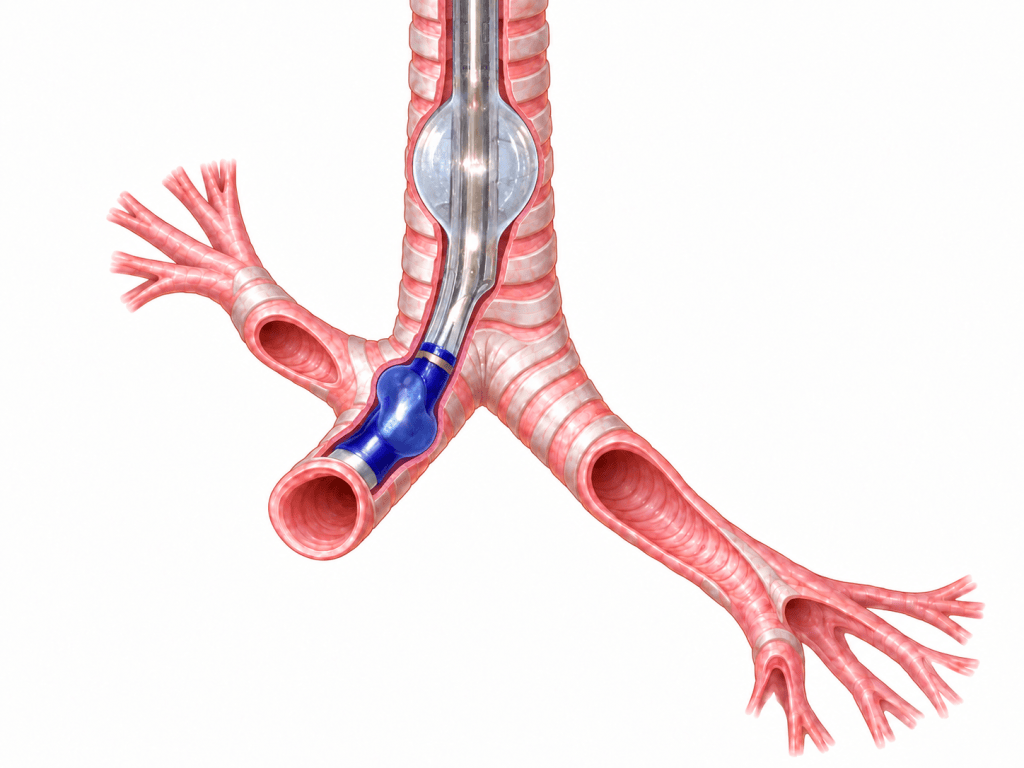
Right-Sided DLT
The right side is considerably less forgiving.
The right upper-lobe bronchus originates very close to the carina.
If a conventional bronchial tube were simply advanced down the right main bronchus it could obstruct the right upper-lobe bronchus while continuing to ventilate the middle and lower lobes.
A right-sided DLT therefore incorporates a special right-upper-lobe ventilation slot.
That slot must align correctly with the opening of the right-upper-lobe bronchus.
The Right Upper Lobe: The Essential Check
Correctly positioning a right-sided DLT requires considerably more than confirming that the tube has entered the right main bronchus.
The practitioner must establish that:
The right upper lobe can still be ventilated.
If the right-upper-lobe ventilation slot does not align correctly with the bronchial opening, the right upper lobe can become partially or completely obstructed even though the middle and lower lobes remain ventilated.
Auscultation over the upper chest is useful, but transmitted breath sounds may give false reassurance.
Flexible bronchoscopy provides the most reliable assessment.
The operator can directly identify the right-upper-lobe bronchial opening and confirm that it corresponds with the ventilation window within the DLT.
The relatively small margin for error is one of the principal reasons right-sided DLTs are more demanding to position.
Tube Size and Selection
DLTs are substantially larger and more rigid than conventional endotracheal tubes.
Common adult sizes include:
32 Fr, 35 Fr, 37 Fr, 39 Fr and 41 Fr.
Traditional tube selection has often been based upon sex and height, using approximate starting points such as:
| Patient | Traditional starting size |
| Smaller adult female | 32–35 Fr |
| Average adult female | 35–37 Fr |
| Smaller adult male | 37–39 Fr |
| Average or larger adult male | 39–41 Fr |
These should be treated as guides rather than rules.
Height and sex do not predict bronchial diameter perfectly.
Two patients of the same height can have very different tracheal and bronchial anatomy.
Where CT imaging is already available, as it often is before thoracic surgery, it can provide valuable information about airway dimensions and unusual anatomy.
If the tube is too large
An oversized tube may increase the risk of:
- difficult passage through the larynx;
- mucosal trauma;
- vocal-cord injury;
- cuff injury;
- bronchial trauma;
- and, rarely, tracheal or bronchial rupture.
If the tube is too small
An undersized tube may:
- require excessive cuff inflation to achieve a seal;
- be less stable;
- migrate more easily;
- provide less reliable lung isolation;
- and permit leakage between the lungs.
The aim is therefore not to use the largest tube that can physically be inserted.
The aim is to select a tube that is appropriate for the patient’s anatomy.
Resistance is information, not an instruction to push harder.
Choosing the Side of the DLT
Tube selection also depends upon where the surgeon intends to operate.
Where anatomy and the planned procedure allow, it is useful to keep the bronchial component of the DLT away from the bronchus that may subsequently need to be divided.
Thoracic operations do not always progress exactly as planned.
A segmentectomy may become a lobectomy.
A lobectomy may occasionally become a more extensive resection.
Tumour position, margins or unexpected pathology can all alter the surgical plan.
If the bronchial tip and cuff are sitting within the bronchus that the surgeon later needs to staple or divide, the airway suddenly becomes part of the operative field.
Or, in traditional theatre language:
Cutting the end off the double-lumen tube usually annoys the anaesthetic team.
The humour makes the point memorable, but the underlying issue is serious.
Before bronchial stapling or division, the surgical and anaesthetic teams should communicate about:
- the position of the bronchial limb;
- the position of the bronchial cuff;
- whether the cuff needs to be deflated;
- whether the tube needs withdrawing;
- and how ventilation will be maintained afterwards.
This is a good example of human factors in thoracic surgery.
The surgeon may be concentrating on margins and vascular anatomy while the anaesthetist is concentrating on oxygenation and lung isolation.
Both need a shared understanding of where the airway device lies.
Two Different Questions
When checking a DLT we are really asking two separate questions.
Is it working?
Can each lung be ventilated independently?
Where exactly is it?
Is the bronchial cuff and distal tube anatomically where we think it is?
Clinical examination helps answer the first question.
Bronchoscopy answers the second.
Both are important.
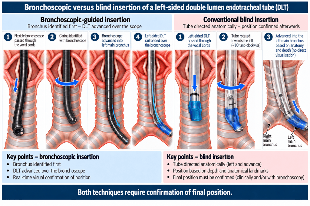
Bronchoscope-Guided Placement
Flexible bronchoscopy can be used not only to confirm final DLT position but also to guide the tube into the intended bronchus.
The principle is simple:
Find the bronchus first, then take the tube to it.
Step 1 – Prepare the Tube and Bronchoscope
The bronchoscope is passed through the appropriate lumen of the DLT so that the tube can later be railroaded over the bronchoscope.
The team should ensure that the following are available and functioning:
- DLT;
- bronchoscope;
- screen or light source;
- suction;
- connector;
- clamps;
- cuff syringes;
- and ventilation equipment.
Step 2 – Pass Between the Vocal Cords
The bronchoscope is advanced between the vocal cords and into the trachea.
The operator advances down the trachea towards the first major landmark:
the carina.
Step 3 – Identify the Anatomy
At the carina, the operator identifies:
the left main bronchus
and
the right main bronchus.
If right-sided placement is intended, the right-upper-lobe bronchus must also be identified.
Step 4 – Advance Into the Intended Bronchus
The bronchoscope is advanced into the selected main bronchus.
The required route has now been anatomically identified.
The operator is no longer estimating which direction the DLT should take.
Step 5 – Railroad the DLT
The DLT is then gently advanced down over the bronchoscope.
The bronchoscope acts as a guide.
However, the bronchoscope is smaller and considerably more flexible than the DLT.
The fact that the scope has passed easily does not mean the larger tube should be forced through resistance.
Step 6 – Confirm the Final Position
The bronchoscope is then used to confirm final positioning.
The operator assesses the relationship between:
- carina;
- bronchial cuff;
- bronchial lumen;
- tracheal lumen;
- and relevant lobar bronchial openings.
For a right-sided DLT, the right-upper-lobe ventilation window must align correctly with the right-upper-lobe bronchus.
The Conventional or “Blind” Technique
Calling this method completely blind can be misleading.
The DLT is usually introduced through the vocal cords using direct or video laryngoscopy.
The “blind” part is the subsequent passage of the bronchial limb from the trachea into the intended main bronchus without directly visualising the bronchial bifurcation.
For a conventional left-sided insertion:
- the DLT is passed through the vocal cords;
- the stylet is removed at the appropriate point;
- the tube is rotated;
- the preformed bronchial tip is directed towards the left;
- the tube is gently advanced;
- expected depth and resistance are assessed;
- cuffs are inflated appropriately;
- lung separation is assessed clinically.
The technique therefore relies upon:
tube curvature + tube rotation + airway anatomy + insertion depth + experience.
The difference between the techniques can be summarised simply:
Blind placement asks the tube to find the bronchus.
Bronchoscopic placement finds the bronchus first and then takes the tube to it.
Does Turning the Head Turn the Carina?
No.
Traditional descriptions of blind DLT placement can sometimes give the impression that turning the patient’s head rotates the carina into a more favourable position.
It does not.
The carina lies within the thorax.
Turning the head primarily changes the orientation of:
- the head;
- the neck;
- the larynx;
- and the cervical trachea.
Head rotation may therefore change the angle at which the tube approaches the lower trachea and bronchi, and this may influence which direction a blindly advanced tube takes.
That is not the same as physically rotating the carina.
The important distinction is:
Turning the head changes the approach of the tube. It does not turn the carina towards the tube.
Rotating the DLT itself is different.
Tube rotation directly changes the orientation of the preformed bronchial tip and therefore has a mechanical effect on where the distal end travels.
Look – Listen – Feel
Clinical confirmation remains useful even when bronchoscopy is available.
A simple teaching framework is:
LOOK – LISTEN – FEEL
LOOK
Observe:
- chest expansion;
- tidal volume;
- capnography;
- tube depth;
- ventilator pressure;
- air leaks;
- and the response when each lumen is isolated.
For a correctly positioned left-sided DLT:
Bronchial lumen ventilated → left lung should expand.
Tracheal lumen ventilated → right lung should expand.
LISTEN
Auscultate both lungs.
Then selectively ventilate each lumen.
Breath sounds should correspond with the lung being ventilated.
However:
Sound travels.
Apparently satisfactory breath sounds do not prove ideal anatomical tube position.
This is one of the limitations of relying solely on clinical assessment.
FEEL
Manual ventilation gives valuable tactile information.
The practitioner may notice changes in:
- compliance;
- resistance;
- reservoir-bag behaviour;
- airway pressure;
- and the transition between two-lung and one-lung ventilation.
Unexpected resistance may indicate:
- tube displacement;
- obstruction;
- cuff problems;
- bronchospasm;
- blood or secretions;
- surgical compression;
- or another mechanical problem.
Modern ventilation technology has not made the anaesthetist’s hand redundant.
Bronchial Cuff Pressure and Minimum Occlusive Volume
The bronchial cuff needs particular care.
Its purpose is to create enough seal to isolate the lung.
It is not designed to be inflated unnecessarily.
The aim is to use the minimum volume and pressure that produce an effective seal.
Bronchial cuff pressures in the region of 20–30 cmH₂O are commonly used as a practical target, with cuff pressure measured using a manometer rather than estimated by squeezing the pilot balloon.
Only small volumes may be required.
A fixed syringe volume should not be treated as a universal rule because cuff design varies considerably between manufacturers.
Too little cuff inflation can cause:
- air leakage;
- incomplete lung separation;
- contamination between lungs;
- poor operative-lung collapse.
Excessive cuff inflation can cause:
- mucosal pressure injury;
- bronchial distortion;
- cuff herniation;
- displacement;
- and potentially severe bronchial injury.
The principle is therefore:
Minimum occlusive volume, not maximum tolerated volume.
Correct Supine Does Not Mean Correct Lateral
One of the most important practical rules in thoracic anaesthesia is:
A DLT confirmed correctly in the supine patient must not automatically be assumed to remain correctly positioned after lateral positioning.
Turning the patient can alter the relationship between:
- tube;
- carina;
- main bronchi;
- head and neck;
- and bronchial cuff.
The practical sequence should therefore be:
Position DLT → confirm → turn patient → reassess.
If there is any doubt, bronchoscopic rechecking should be performed.
Suction Through a DLT
One major advantage of a DLT is that each lung can be accessed separately.
A suction catheter can be passed through either lumen, allowing blood and secretions to be removed selectively.
This is particularly valuable in:
- pulmonary haemorrhage;
- infection;
- major secretions;
- or contamination restricted predominantly to one lung.
However, DLT lumens are narrower than an equivalent conventional single-lumen tube.
This means:
- suction catheter size matters;
- thick secretions may occlude a lumen;
- blood clot can cause major obstruction;
- and apparent tube malposition may actually be intraluminal blockage.
Difficulty passing the expected suction catheter is itself useful diagnostic information.
What Bronchoscopy Can Get Wrong
Flexible bronchoscopy is the reference standard for confirming anatomical DLT position.
It is not infallible.
Problems include:
Blood
Blood may rapidly obscure the distal lens.
Secretions
Mucus can make landmarks difficult or impossible to identify.
Fogging and contamination
The image may repeatedly deteriorate despite suction and cleaning.
Distorted anatomy
Tumour, previous surgery, trauma or congenital anatomy may make the airway look very different from textbook illustrations.
Misidentification of anatomy
An inexperienced bronchoscopist can misinterpret distal bronchial anatomy or mistake a more distal bifurcation for the carina.
Tube movement after confirmation
A perfect image only confirms the position at that moment.
The tube can move afterwards.
The bronchoscope fits where the DLT will not
A thin flexible scope may negotiate an angle that a large, relatively rigid DLT cannot safely follow.
Therefore:
Bronchoscopy provides direct anatomical information, but the image still has to be visible and correctly interpreted.
Traditional Direct-View Bronchoscopy Versus Video Bronchoscopy
Traditionally, flexible bronchoscopy involved the anaesthetist looking directly through the eyepiece of the bronchoscope.
This is effective, but it creates a single-observer system.
One person sees the airway.
Everyone else depends on that person’s verbal description.
Modern disposable video bronchoscopes offer a significant advantage because the image is displayed on a screen.
Several members of the theatre team can observe the bronchoscopy simultaneously.
The anaesthetist remains responsible for performing and interpreting the procedure, but the ODP, anaesthetic nurse, trainee and, where appropriate, surgeon can also see:
- vocal cords;
- trachea;
- carina;
- left and right main bronchi;
- bronchial cuff;
- DLT position;
- blood;
- secretions;
- obstruction;
- and the right-upper-lobe bronchial opening where relevant.
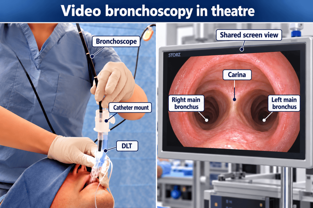
Shared Anatomical Confirmation
When several experienced practitioners can see the same image, anatomy can be discussed in real time.
For example:
“That is the carina.”
“We are entering the left main bronchus.”
“The bronchial cuff is just below the carina.”
“The right-upper-lobe opening is visible.”
This provides an opportunity for another experienced practitioner to confirm or question what is being seen.
The purpose is not to undermine the person controlling the bronchoscope.
It is to reduce the risk of a single-observer error.
Anticipating Clinical Need
The greatest benefit for the ODP or anaesthetic nurse may be the ability to anticipate rather than simply react.
If the practitioner can see that:
- secretions are obscuring the view;
- the cuff is too proximal;
- the tube is too deep;
- the tube needs withdrawing;
- suction is required;
- the anatomy is difficult to interpret;
- or the current strategy is failing,
they can prepare what is likely to be needed next.
This may include:
- suction;
- cuff syringe;
- cuff manometer;
- clamps;
- tube fixation;
- another bronchoscope;
- another DLT;
- or equipment for an alternative airway strategy.
The practitioner moves from being a passive assistant to becoming an active member of the airway-management team.
Video Bronchoscopy and Human Factors
Video bronchoscopy therefore offers more than simply a different way of viewing the airway.
It provides a major human-factors benefit.
Traditional direct-view bronchoscopy creates a single-person information channel.
Video bronchoscopy creates a shared visual information channel.
Several people can see the same airway at the same time.
This may improve:
- communication;
- situational awareness;
- anatomical agreement;
- teaching;
- anticipation of equipment needs;
- troubleshooting;
- and team decision-making.
The difference can be summarised simply:
Traditional bronchoscopy
One person sees the airway and tells the team what they can see.
Video bronchoscopy
The team sees the airway together.
This supports one of the central principles of good human-factors practice:
Create a shared mental model of what is happening now and what is likely to happen next.
Why Trauma Is Different
Elective thoracic anaesthesia generally provides:
- a planned operation;
- prepared equipment;
- a relatively controlled airway;
- established anatomy;
- and time to optimise tube position.
Major trauma may provide:
- blood;
- vomit;
- secretions;
- clot;
- haemodynamic instability;
- hypoxia;
- distorted anatomy;
- haemothorax;
- pulmonary injury;
- or a rapidly progressing thoracotomy.
A theoretical gold-standard bronchoscopic view is of little immediate use if the lens is repeatedly covered in blood.
This is where practical clinical airway assessment remains important.
Why the Left DLT Is Particularly Attractive in Trauma
A right-sided DLT requires accurate alignment between the right-upper-lobe ventilation slot and the patient’s right-upper-lobe bronchus.
That requires greater positional precision.
If bronchoscopic visualisation is compromised by blood or contamination, identifying those landmarks may be extremely difficult.
The left main bronchus provides a larger margin for safe placement and does not require alignment with an early upper-lobe bronchial opening.
Where a DLT is appropriate and there is no contraindication to occupying the left main bronchus, the left-sided DLT therefore offers more forgiving anatomy.
Emergency Anatomical Placement
In severe trauma, priorities may initially be:
Airway → Oxygenation → Ventilation → Lung separation → Surgical control.
If bronchoscopic visualisation cannot initially be achieved, an experienced anaesthetist may have to establish anatomical placement and assess immediate function using:
Look – Listen – Feel.
The immediate questions become:
- Can the patient be ventilated?
- Has lung separation been achieved?
- Is the unaffected lung protected?
- Is oxygenation adequate?
- Can surgery proceed?
Bronchoscopy can then be used when conditions and visibility allow.
This is not an argument that blind placement is superior.
It recognises that in emergency care:
Function may occasionally have to be established before perfect anatomical confirmation is possible.
When a DLT May Not Be the Best Choice
A DLT is extremely useful, but it is not automatically the correct device for every patient.
Alternative strategies should be considered in situations including:
Known or anticipated difficult airway
A bulky DLT may be significantly more difficult to introduce than a conventional single-lumen tube.
Securing the airway may take priority over immediate lung isolation.
Significant tracheal or bronchial trauma
A relatively rigid DLT advanced through a damaged airway may worsen the injury or create a false passage.
Very small adult airway
A smaller DLT or conventional single-lumen tube with bronchial blocker may be more appropriate.
Severely abnormal bronchial anatomy
Previous surgery, tumour or congenital anatomy may prevent conventional DLT positioning.
Existing tracheostomy
A standard oral DLT may be inappropriate.
A patient already safely intubated
Particularly in trauma, replacing a secure airway simply to insert a DLT requires careful consideration.
Tube Exchange: Do Not Lose a Good Airway Lightly
Consider a trauma patient arriving in theatre with:
- a secure single-lumen endotracheal tube;
- satisfactory capnography;
- acceptable ventilation;
- and reasonable oxygenation.
Changing that tube for a DLT is not a trivial administrative step.
It means deliberately removing a functioning airway and replacing it with a larger, more complex device.
Potential risks include:
- failure to re-intubate;
- airway trauma;
- loss of oxygenation;
- aspiration;
- haemodynamic deterioration;
- and complete loss of an airway that was previously secure.
This is particularly important in:
- facial trauma;
- cervical instability;
- airway oedema;
- massive haemorrhage;
- or known difficult intubation.
A bronchial blocker passed through the existing single-lumen tube may sometimes provide lung isolation without sacrificing the established airway.
The question should therefore be:
What is the safest way to isolate this patient’s lung?
rather than:
Thoracic surgery means we must insert a DLT.
Loss of One-Lung Ventilation: A Practical ODP Sequence
When apparently successful one-lung ventilation suddenly deteriorates, a systematic approach is more useful than random adjustments.
A practical sequence is:
1. CIRCUIT
Check the obvious first.
- Is the circuit connected?
- Is the correct lumen connected?
- Are clamps correctly positioned?
- Has a connector become loose?
- Is there an obvious leak?
- Is capnography present?
2. TUBE
Look at the DLT.
- Has the depth at the teeth changed?
- Has the tube rotated?
- Has the head recently moved?
- Has the patient just been turned?
- Have the cuffs changed?
3. VENTILATION
Look, Listen and Feel.
- Which lung is moving?
- Where are breath sounds heard?
- Has airway pressure increased?
- Has compliance changed?
- Has tidal volume fallen?
4. SUCTION / OBSTRUCTION
Consider:
- mucus;
- secretions;
- blood;
- clot;
- kinked tubing;
- or obstruction of a lumen.
Attempt appropriate suction.
Difficulty passing the suction catheter may itself identify the problem.
5. BRONCHOSCOPY
If the cause is not obvious, inspect:
- tube depth;
- cuff position;
- bronchial openings;
- obstruction;
- blood;
- secretions;
- and the relationship between tube and carina.
6. SURGERY
Do not forget the person operating inside the chest.
Ask:
- Is the surgeon manipulating the bronchus?
- Has the hilum been mobilised?
- Is the bronchus being compressed?
- Has a clamp been placed?
- Has lung retraction changed?
- Has the operation progressed to a different resection?
The answer is not always at the anaesthetic machine.
A useful mental prompt is:
Circuit → Tube → Ventilation → Suction → Scope → Surgeon.
Hypoxaemia During One-Lung Ventilation
A falling oxygen saturation during OLV should always prompt assessment, but it does not automatically mean that the DLT has moved.
One-lung ventilation deliberately creates an intrapulmonary shunt because the non-ventilated lung continues to receive some blood flow.
Hypoxic pulmonary vasoconstriction reduces perfusion to that lung, but it does not remove the shunt completely.
Potential causes of hypoxaemia include:
- DLT malposition;
- obstruction by blood or secretions;
- atelectasis in the dependent ventilated lung;
- inadequate ventilation;
- excessive or inadequate PEEP;
- significant shunt through the operative lung;
- low cardiac output;
- anaemia;
- pre-existing lung disease;
- dependent-lung pneumothorax;
- bronchospasm;
- surgical compression;
- and altered hypoxic pulmonary vasoconstriction.
The practical message is:
Desaturation means investigate, not automatically advance or withdraw the DLT.
Severe hypoxaemia may require temporary return to two-lung ventilation while the cause is identified.
Possible Complications
DLTs are generally safe in experienced hands, but they are larger and more rigid than conventional endotracheal tubes and carry specific risks.
Minor airway trauma
- sore throat;
- mucosal injury;
- hoarseness;
- minor bleeding.
Laryngeal injury
- vocal-cord trauma;
- arytenoid injury;
- cuff-related damage.
Tracheal or bronchial injury
Rarely:
- bronchial tear;
- tracheal rupture;
- pneumothorax;
- pneumomediastinum;
- surgical emphysema.
Malposition
One of the commonest practical problems.
Obstruction
Blood, secretions and mucus may block one lumen.
Inadequate isolation
Poor cuff seal or malposition may allow continued ventilation of the operative lung.
Hypoxaemia
This may result from tube problems or from the physiology of one-lung ventilation itself.
The general rule remains:
If something does not feel right, do not simply apply more force or more pressure. Find out why.
End of Surgery: What Happens to the DLT?
A DLT is primarily a lung-isolation device.
It is not usually the ideal tube for prolonged postoperative ventilation.
Reasons include:
- large external diameter;
- narrow individual lumens;
- increased resistance;
- difficulty clearing secretions;
- cuff-related concerns;
- and risk of displacement.
If the patient can be extubated safely at the end of surgery, the DLT is removed in the normal way.
If postoperative ventilation is required, the DLT may need to be exchanged for a conventional single-lumen tube.
This exchange should be planned.
Questions include:
- Was intubation difficult?
- Is airway oedema expected?
- Has the airway been surgically manipulated?
- Has there been major fluid resuscitation?
- Would an airway-exchange catheter be appropriate?
- What would happen if the airway were lost now?
Postoperative tube exchange should therefore not be regarded as routine housekeeping.
It is another airway-management procedure.
The ODP and Anaesthetic Nurse Role
DLT management demonstrates why an anaesthetic assistant should understand the procedure rather than simply know where the equipment is stored.
Before induction the practitioner should anticipate requirements for:
- appropriate DLT sizes;
- left- and right-sided tubes where appropriate;
- direct or video laryngoscopy;
- stylet;
- flexible bronchoscope;
- functioning video display;
- suction;
- appropriate suction catheters;
- cuff manometer;
- clamps;
- connectors;
- syringes;
- bronchial blocker;
- airway-exchange equipment;
- and conventional single-lumen tubes.
The ODP should understand what the bronchoscopist is attempting to identify.
When the anaesthetist says:
“Carina.”
the practitioner should understand the landmark.
When a right-sided tube is being positioned, they should understand why:
“Can I see the right upper lobe?”
is a critical question.
When one-lung ventilation suddenly deteriorates, they should already be thinking:
Circuit → Tube → Ventilation → Suction → Scope → Surgeon.
That is the difference between simply assisting with an airway procedure and actively contributing to safe thoracic anaesthesia.
Bronchoscopy Versus Look, Listen and Feel
These techniques should not be presented as:
modern versus obsolete
or
correct versus incorrect.
They answer different questions.
Bronchoscopy tells us:
Where is the tube?
Look, Listen and Feel tells us:
What are the lungs actually doing?
A beautifully positioned DLT is of little value if the patient cannot be ventilated effectively.
Equally, apparently satisfactory breath sounds do not prove that the tube is optimally positioned.
The techniques are therefore complementary.
Elective Thoracic Surgery Versus Major Trauma
| Elective Thoracic Surgery | Major Thoracic Trauma |
| Planned airway strategy | Airway strategy may evolve rapidly |
| Controlled airway | Blood and contamination may be present |
| Anatomy generally intact | Anatomy may be disrupted |
| Bronchoscope readily available | Bronchoscopic view may be very poor |
| Time to optimise position | Immediate lung separation may be required |
| Direct anatomical confirmation expected | Functional confirmation may initially be necessary |
| Right or left tube chosen according to anatomy and surgery | Left DLT often offers more forgiving anatomy when appropriate |
| Recheck after lateral positioning | Reassessment may need to be continuous |
| Planned extubation or exchange strategy | Existing secure airway may be more valuable than ideal lung-isolation equipment |
The objective is the same:
safe and effective lung isolation.
The route used to achieve it may be very different.
Key Principles for Nurses and ODPs
A left-sided DLT is generally more forgiving than a right-sided tube.
Correct positioning of a right-sided DLT requires confirmation that the right upper lobe remains ventilated.
Tube size should reflect airway anatomy rather than sex or height alone.
A larger tube is not automatically a better tube.
Use the minimum bronchial cuff volume and pressure required to obtain a seal.
Correct supine does not mean correct lateral.
The surgical procedure influences where the bronchial limb should lie.
Flexible bronchoscopy remains the reference standard for confirming anatomical DLT position.
Bronchoscopy is only useful when the image can be seen and correctly interpreted.
Video bronchoscopy creates a shared visual field and improves team situational awareness.
A bronchoscope can also guide a DLT into an identified bronchus.
Blind bronchial placement relies upon tube orientation, anatomy, depth and subsequent clinical assessment.
Turning the patient’s head does not rotate the carina.
Look, Listen and Feel provides valuable functional information but does not prove exact anatomical position.
Blood and secretions may make bronchoscopic identification extremely difficult during trauma.
A secure single-lumen airway should not automatically be sacrificed simply because lung isolation is required.
Hypoxaemia during OLV does not automatically mean DLT malposition.
Sudden deterioration requires systematic investigation rather than random tube manipulation.
DLTs are not usually ideal for prolonged postoperative ventilation.
And perhaps the most useful distinction to remember is:
Blind placement asks the tube to find the bronchus. Bronchoscopic placement finds the bronchus first and then takes the tube to it.
For the Operating Department Practitioner or anaesthetic nurse, understanding these principles transforms DLT management from simply handing an anaesthetist an unusual airway device into understanding what the team is trying to achieve, recognising when it has succeeded and, most importantly, recognising when something has changed.

