Understanding neuromonitoring, spinal cord perfusion and the role of the theatre team
Thoracoabdominal aortic surgery is amongst the most complex procedures undertaken within the operating theatre.
Although the primary objective is to repair a diseased or dissected section of the aorta, one of the most significant risks associated with surgery involving the descending thoracic and abdominal aorta is spinal cord ischaemia.
If the blood supply to the spinal cord becomes inadequate, motor pathways can become progressively dysfunctional and, if circulation is not restored, irreversible neurological injury may occur.
The consequence may be weakness, paraparesis or permanent paraplegia.
For nurses and Operating Department Practitioners working within major vascular, cardiac or cardiothoracic theatres, understanding why the spinal cord is at risk, how it is monitored and what happens when monitoring deteriorates is therefore an important part of understanding the procedure as a whole.
Basic Spinal Anatomy
The spinal cord runs within the vertebral canal and forms the principal communication pathway between the brain and the peripheral nervous system.
It is protected by:
- the vertebral column;
- the meninges;
- cerebrospinal fluid;
- surrounding muscles and connective tissues.
Spinal nerve roots emerge from the cord and leave the vertebral canal through the intervertebral foramina before forming peripheral nerves.
The spinal cord itself is divided broadly into cervical, thoracic, lumbar and sacral regions, although the spinal cord terminates above the lower lumbar vertebrae and continues distally as the cauda equina.
For theatre practitioners, the important point is that the spinal cord is not simply a cable running down the back.
It is metabolically active neurological tissue that is extremely dependent upon continuous oxygenated blood flow.
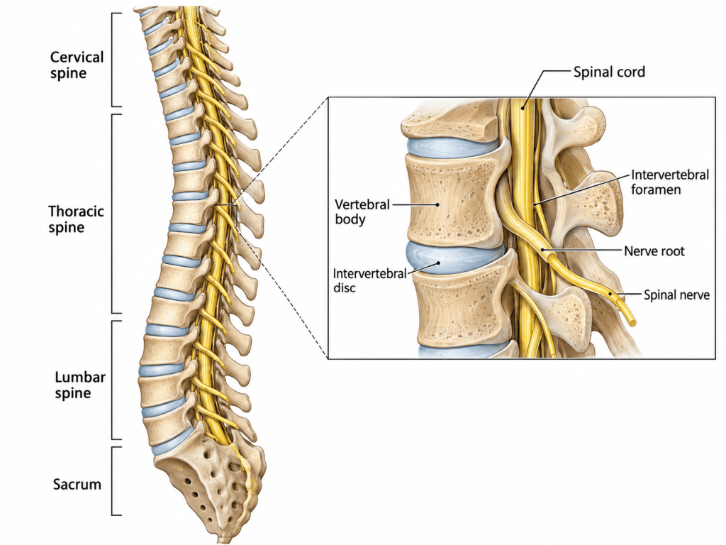
Spinal anatomy showing the cervical, thoracic and lumbar spine, spinal cord, nerve roots and spinal nerves.
What Is Spinal Cord Stimulation?
Electrical stimulation of the nervous system is used in several different clinical settings.
One example is the implanted spinal cord stimulator, used primarily for the treatment of chronic neuropathic pain.
A conventional spinal cord stimulation system consists of:
- one or more epidural electrode leads;
- electrical contacts positioned close to the spinal cord;
- connecting wires;
- an implantable pulse generator;
- a patient controller or programming system.
The implantable pulse generator delivers electrical impulses through the epidural electrodes.
These impulses modify neurological signalling and can alter the perception of chronic pain.
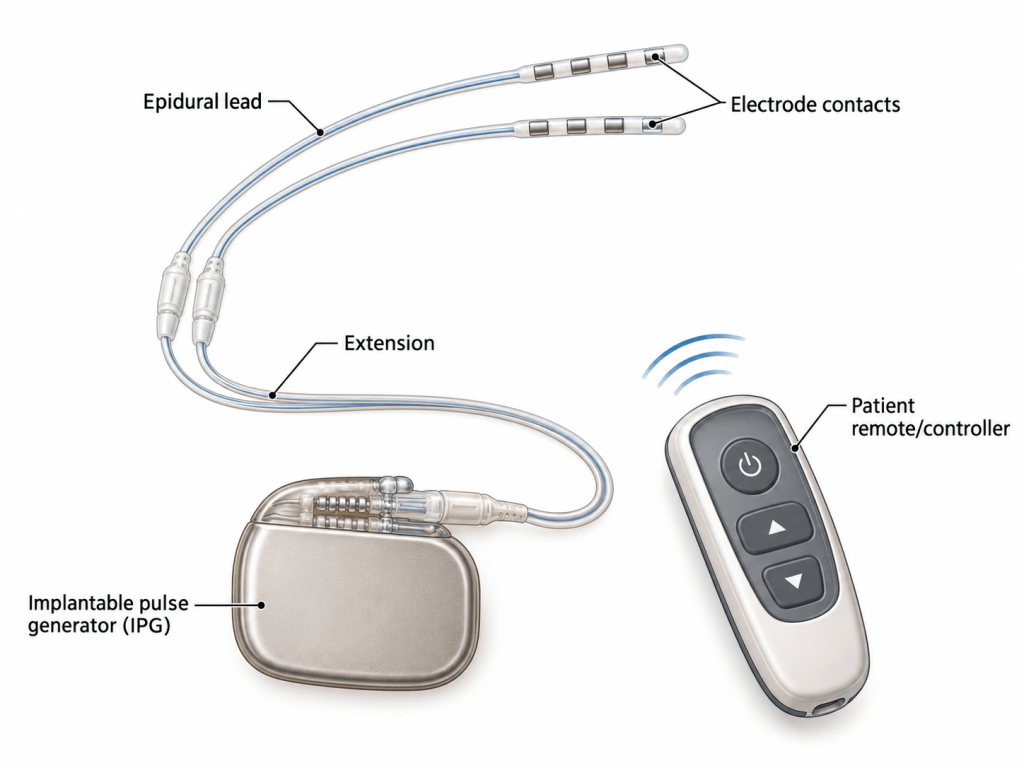
Components of an implanted spinal cord stimulator: epidural leads, electrode contacts, extension cable, pulse generator and patient controller.
The leads are normally positioned within the epidural space, outside the dura surrounding the spinal cord.
The pulse generator is usually implanted subcutaneously in the flank, abdomen or upper buttock region.
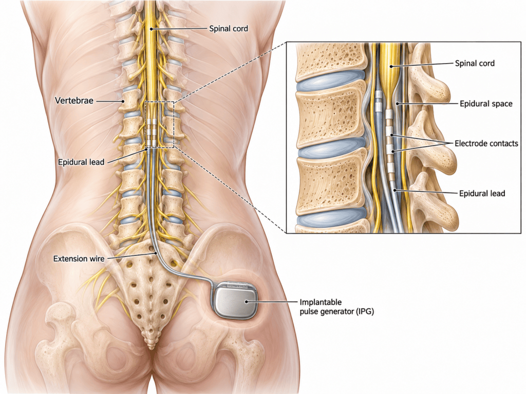
Spinal anatomy showing the location of spinal cord stimulator leads within the epidural space and the subcutaneous pulse generator.
However, this is not the same as the electrical stimulation used during thoracoabdominal aortic surgery.
This distinction is important.
An implanted spinal cord stimulator provides therapy.
Intraoperative neurophysiological stimulation provides information.
During thoracoabdominal aortic surgery, electrical stimulation may be used to determine whether the motor pathways running through the spinal cord remain functional.
Why Is the Spinal Cord at Risk During Aortic Surgery?
The spinal cord receives its blood supply from a complex network of vessels.
The major longitudinal vessels include:
- the anterior spinal artery;
- the paired posterior spinal arteries.
These vessels are reinforced by segmental arteries entering the spinal circulation at different levels.
Many of these originate indirectly from the:
- intercostal arteries;
- lumbar arteries;
- branches of the thoracic and abdominal aorta.
One particularly important vessel is the great anterior radiculomedullary artery, more commonly known as the artery of Adamkiewicz.
Its origin varies considerably between individuals.
This anatomical variability is important because the blood supply to the spinal cord does not depend upon one identical arterial arrangement in every patient.
Instead, spinal cord perfusion is maintained by a complex collateral vascular network.
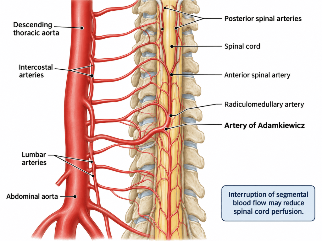
Simplified arterial supply to the spinal cord showing the thoracic and abdominal aorta, intercostal and lumbar arteries, anterior spinal artery and artery of Adamkiewicz.
Thoracoabdominal Aortic Dissection
An aortic dissection occurs when blood enters the wall of the aorta through a tear in the intima.
Blood then tracks between layers of the aortic wall, creating a false lumen alongside the original true lumen.
When a dissection extends through the descending thoracic aorta and into the abdominal aorta, blood flow into vessels originating from the affected segment may become compromised.
Surgical repair may require sections of the diseased aorta to be:
- clamped;
- opened;
- replaced;
- bypassed;
- reconstructed with a vascular graft.
During this process, intercostal and lumbar arteries supplying the spinal collateral network may temporarily or permanently lose their normal blood supply.
The spinal cord may consequently become ischaemic.
Spinal Cord Perfusion
The spinal cord requires a sufficient pressure gradient to maintain blood flow.
A useful simplified relationship is:
Spinal Cord Perfusion Pressure ≈ Mean Arterial Pressure − Cerebrospinal Fluid Pressure
This means spinal cord perfusion can deteriorate in two important ways.
Mean arterial pressure may fall.
Or cerebrospinal fluid pressure may rise.
In practice, both may occur simultaneously.
Other factors can further compromise oxygen delivery, including:
- anaemia;
- hypoxaemia;
- reduced cardiac output;
- major haemorrhage;
- prolonged aortic cross-clamping;
- loss of important segmental arteries.
Maintaining spinal cord perfusion is therefore not dependent upon one intervention.
It requires a coordinated strategy.
Cerebrospinal Fluid Drainage
A lumbar cerebrospinal fluid drain may be inserted before major thoracoabdominal aortic surgery.
The catheter enters the intrathecal space and allows controlled drainage of cerebrospinal fluid.
Reducing CSF pressure can increase the pressure gradient available to perfuse the spinal cord.
For an ODP or theatre nurse, the CSF drain should not be regarded simply as another drain.
It forms part of the patient’s neurological protection strategy.
Careful attention is required to:
- drainage height;
- CSF pressure;
- drainage volume;
- catheter security;
- sterile connections;
- prescribed drainage limits.
Excessive drainage is potentially dangerous.
The drainage system must therefore be managed according to local protocol and specialist instructions.
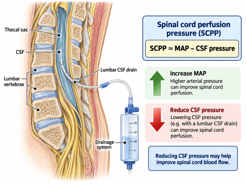
Lumbar CSF drain showing catheter position, drainage system and the relationship between MAP, CSF pressure and spinal cord perfusion.
How Do We Know Whether the Spinal Cord Is Still Working?
One of the difficulties during general anaesthesia is that the patient cannot tell us whether their legs have become weak.
By the time the patient wakes, irreversible neurological damage may already have occurred.
This is where intraoperative neurophysiological monitoring becomes valuable.
One of the most important techniques used during thoracoabdominal aortic surgery is the monitoring of:
Motor-Evoked Potentials — MEPs
MEPs assess the functional integrity of the motor pathways.
An electrical stimulus is applied through electrodes positioned over the scalp.
The stimulus activates the motor cortex.
The resulting electrical activity travels:
Motor cortex
↓
Corticospinal tract
↓
Spinal cord
↓
Motor neurones
↓
Peripheral nerves
↓
Skeletal muscle
Recording electrodes positioned over muscles in the legs detect the resulting muscular electrical response.
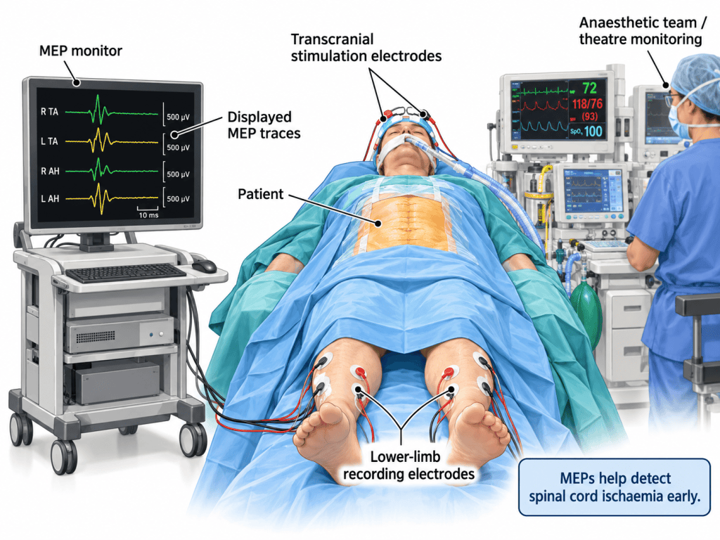
Theatre setup for MEP monitoring showing transcranial stimulation electrodes, lower-limb recording electrodes and the MEP monitoring system.
What Does the MEP Monitor Tell Us?
MEP waveforms provide the neurophysiology and theatre teams with information about whether the motor pathways remain electrically functional.
At the beginning of the procedure, baseline responses are established.
These become the reference against which subsequent signals are compared.
A stable MEP response suggests that the motor pathway is functioning.
A significant reduction in amplitude or complete disappearance of the response may suggest deterioration.
It is important, however, to remember that a falling MEP is a warning, not a diagnosis.
Changes can occur because of:
- spinal cord ischaemia;
- hypotension;
- anaesthetic agents;
- neuromuscular blockade;
- hypothermia;
- technical problems;
- displaced electrodes.
The significance of any change therefore has to be interpreted in the context of what is happening to the patient at that moment.
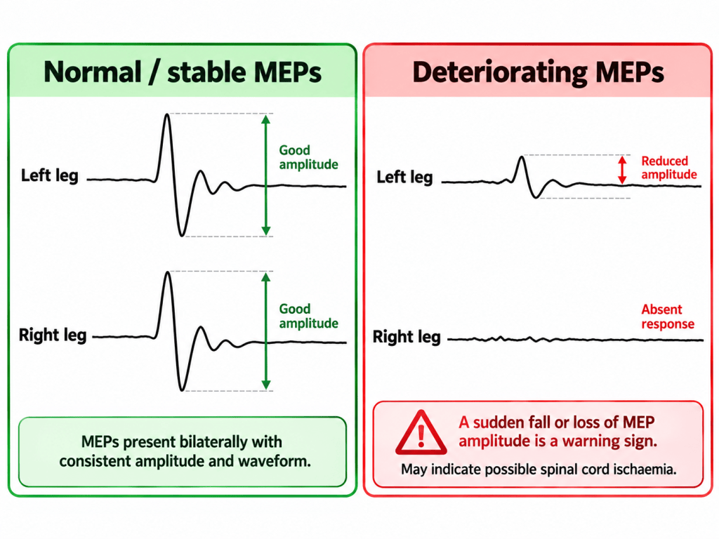
Comparison between normal/stable MEP waveforms and deteriorating or absent MEP responses.
Anaesthesia and MEP Monitoring
MEP monitoring has important implications for the anaesthetic technique.
Because the final signal depends upon a muscle contracting in response to stimulation, profound neuromuscular blockade may prevent useful MEP recordings.
The anaesthetic team must therefore balance surgical requirements against the need to preserve reliable neuromonitoring.
Volatile anaesthetic agents can also suppress evoked potentials.
Total intravenous anaesthesia may therefore be used in cases where reliable MEP monitoring is required.
Temperature, blood pressure, oxygenation and haemoglobin concentration also influence neurological function and signal quality.
For the anaesthetic practitioner or ODP, this means an unexpected MEP change may immediately prompt consideration of:
- anaesthetic depth;
- neuromuscular blockade;
- arterial pressure;
- haemoglobin;
- oxygenation;
- ventilation;
- temperature;
- equipment integrity.
What Happens When the MEPs Fall?
A significant reduction or disappearance of MEPs requires immediate communication.
The most important action is:
Tell the team.
The neurophysiologist, anaesthetist, surgeon, perfusionist and theatre team must rapidly determine whether the change represents a technical problem or genuine deterioration in spinal cord perfusion.
The response may include:
- confirming electrode connections;
- checking whether the change is unilateral or bilateral;
- increasing mean arterial pressure;
- reviewing oxygenation and ventilation;
- checking haemoglobin and blood loss;
- reviewing distal aortic perfusion;
- checking bypass or pump flows;
- reviewing CSF pressure and drainage;
- considering the position of the aortic clamp;
- restoring blood flow to important segmental arteries.
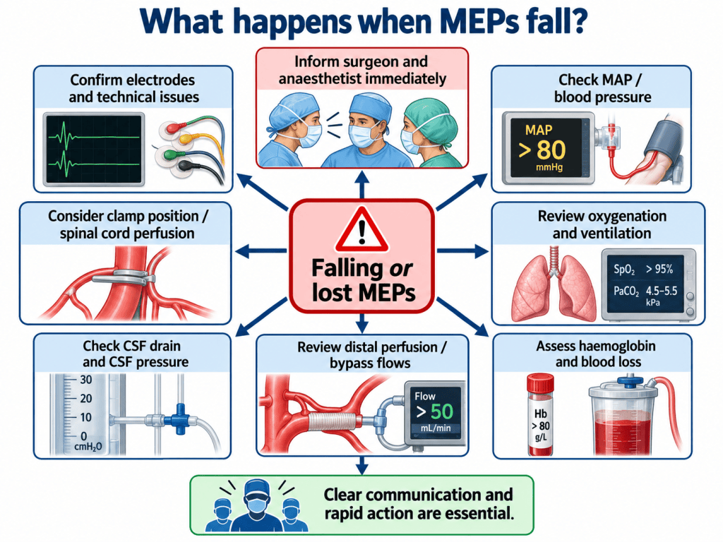
“What happens when MEPs fall?” team response infographic.
This is where the role of the wider theatre team becomes particularly important.
An ODP may not decide whether an intercostal artery should be reimplanted.
A theatre nurse may not determine the required distal perfusion pressure.
But both need to understand why the team suddenly changes direction when neuromonitoring deteriorates.
Sequential Aortic Clamping
During extensive thoracoabdominal reconstruction, sections of the aorta may be dealt with sequentially.
This can allow the surgical team to observe what happens to spinal cord function as different regions of the aorta are excluded.
If the MEPs remain stable following clamping of a particular segment, spinal cord perfusion may remain adequate through collateral circulation.
If the MEPs deteriorate shortly after a clamp is applied, interruption of an important part of the collateral network must be considered.
This may influence subsequent surgical and perfusion decisions.
MEPs therefore do considerably more than simply monitor the patient.
They can contribute directly to surgical decision-making.
Why Can Paraplegia Occur After Surgery?
Spinal cord injury does not necessarily occur during the operation itself.
A patient may wake from surgery moving both legs normally and deteriorate several hours later.
Delayed spinal cord ischaemia may be associated with:
- postoperative hypotension;
- bleeding;
- anaemia;
- reduced cardiac output;
- increased CSF pressure;
- deterioration of collateral blood flow.
This is why spinal cord protection continues into postoperative intensive care.
Blood pressure targets and CSF drainage protocols may continue for a considerable period after surgery.
The handover from theatre to critical care must therefore clearly communicate:
- neurological monitoring;
- CSF drain instructions;
- blood pressure targets;
- haemoglobin considerations;
- intraoperative MEP changes;
- interventions performed in response.
MEPs and SSEPs
Another form of neurophysiological monitoring sometimes used alongside MEPs is the somatosensory-evoked potential, or SSEP.
SSEPs assess sensory rather than motor pathways.
A peripheral nerve, commonly in the lower limb, is electrically stimulated and the resulting signal is detected further along the nervous system.
MEPs and SSEPs therefore assess different neurological pathways.
For the theatre practitioner, the simple distinction is:
MEPs ask: “Can the brain still communicate with the muscles?”
SSEPs ask: “Can sensory information still travel towards the brain?”
Using more than one monitoring modality may provide additional information about spinal cord function.
The Theatre Team
Thoracoabdominal aortic surgery demonstrates particularly clearly why major surgery depends upon teamwork.
Spinal cord protection may involve:
- surgeon;
- anaesthetist;
- anaesthetic practitioner or ODP;
- scrub practitioner;
- circulating practitioner;
- perfusionist;
- neurophysiologist;
- radiographer;
- intensive-care team.
Each professional sees the operation from a slightly different perspective.
The neurophysiologist sees the disappearing MEP.
The anaesthetist sees the falling arterial pressure.
The perfusionist sees the falling distal flow.
The surgeon knows that a new aortic segment has just been clamped.
The ODP may simultaneously recognise a change in anaesthetic conditions or prepare the equipment needed for the next intervention.
Individually, these pieces of information may appear unrelated.
Together, they may identify developing spinal cord ischaemia.
Human Factors
A monitor is only useful if somebody responds to it.
This is perhaps one of the most important lessons of intraoperative neuromonitoring.
A technically perfect MEP monitoring system cannot prevent neurological injury by itself.
What matters is the team’s response.
A statement as simple as:
“We have lost the MEPs in both legs.”
should immediately create a shared mental model.
Something has changed.
The spinal cord may be at risk.
The cause must be identified.
Interventions may need to occur simultaneously rather than sequentially.
Clear communication therefore becomes part of spinal cord protection.
What Should the ODP or Theatre Nurse Understand?
You do not need to be a vascular surgeon or neurophysiologist to understand what is happening.
A useful practical framework is:
Before surgery
Know:
- whether MEP monitoring is planned;
- whether a CSF drain is present;
- whether distal perfusion or bypass will be used;
- where the monitoring equipment is positioned;
- what equipment may be required urgently.
During surgery
Be aware of:
- major changes in arterial pressure;
- significant blood loss;
- changes in bypass or perfusion;
- movement of monitoring electrodes;
- changes in anaesthetic technique;
- announcements from neurophysiology.
When MEPs deteriorate
Expect:
- immediate communication;
- rapid haemodynamic review;
- equipment checks;
- possible changes to perfusion;
- CSF drain assessment;
- possible changes in surgical strategy.
At handover
Ensure that important information accompanies the patient, particularly:
- intraoperative neurological monitoring events;
- current neurological status where assessable;
- CSF drainage instructions;
- blood pressure targets;
- major transfusion or haemoglobin concerns;
- significant changes in spinal cord monitoring.
Conclusion
Thoracoabdominal aortic dissection surgery places the spinal cord at particular risk because the operation can interrupt the complex arterial network that maintains spinal cord perfusion.
Modern spinal cord protection therefore relies upon several strategies working together.
These may include:
- maintaining adequate arterial pressure;
- preserving oxygen delivery;
- distal aortic perfusion;
- CSF drainage;
- careful management of segmental arteries;
- MEP monitoring;
- rapid multidisciplinary communication.
Electrical stimulation during the procedure does not itself protect the spinal cord.
Instead, motor-evoked potentials provide the theatre team with an early warning that the motor pathways may be deteriorating.
This creates an opportunity to intervene before neurological damage becomes irreversible.
For the nurse or ODP, the important question is therefore not simply:
“What is that monitor doing?”
It is:
“What is the monitor telling the team about the spinal cord — and what happens next if that signal disappears?”
Understanding that relationship turns spinal cord monitoring from an unfamiliar collection of wires, electrodes and waveforms into something much more meaningful:
a real-time warning system protecting one of the patient’s most vulnerable neurological structures.

